
Likes
25
Share
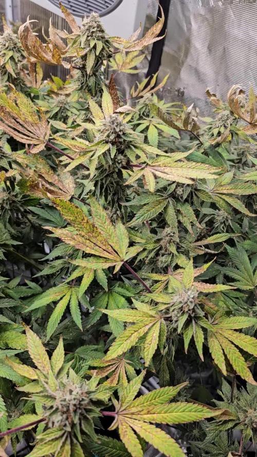
ANTHOCYANIN production is primarily controlled by the Cryptochrome (CR1) Photoreceptor ( !! UV and Blue Spectrums are primary drivers in the production of the pigment that replaces chlorophyll, isn't that awesome! 1. Diverse photoreceptors in plants Many civilizations, including the sun god of ancient Egypt, thought that the blessings of sunlight were the source of life. In fact, the survival of all life, including humans, is supported by the photosynthesis of plants that capture solar energy. Plants that perform photosynthesis have no means of transportation except for some algae. Therefore, it is necessary to monitor various changes in the external environment and respond appropriately to the place to survive. Among various environmental information, light is especially important information for plants that perform photosynthesis. In the process of evolution, plants acquired phytochrome, which mainly receives light in the red light region, and multiple blue light receptors, including his hytropin and phototropin, in order to sense the light environment. .. In addition to these, an ultraviolet light receptor named UVR8 was recently discovered. The latest image of the molecular structure and function of these various plant photoreceptors (Fig. 1), focusing on phytochrome and phototropin. Figure 1 Ultraviolet-visible absorption spectra of phytochrome, cryptochrome, phototropin, and UVR8. The dashed line represents each bioactive absorption spectrum. 2. Phytochrome; red-far red photoreversible molecular switch What is phytochrome? Phytochrome is a photochromic photoreceptor, and has two absorption types, a red light absorption type Pr (absorption maximum wavelength of about 665 nm) and a far-red light absorption type Pfr (730 nm). Reversible light conversion between the two by red light and far-red light, respectively(Fig. 1A, solid line and broken line). In general, Pfr is the active form that causes a physiological response. With some exceptions, phytochrome can be said to function as a photoreversible molecular switch. The background of the discovery is as follows. There are some types of plants that require light for germination (light seed germination). From that study, it was found that germination was induced by red light, the effect was inhibited by subsequent far-red light irradiation, and this could be repeated, and the existence of photoreceptors that reversibly photoconvert was predicted. In 1959, its existence was confirmed by the absorption spectrum measurement of the yellow sprout tissue, and it was named phytochrome. Why does the plant have a sensor to distinguish between such red light and far-red light? There is no big difference between the red and far-red light regions in the open-field spectrum of sunlight, but the proportion of red light is greatly reduced due to the absorption of chloroplasts in the shade of plants. Similar changes in light quality occur in the evening sunlight. Plants perceive this difference in light quality as the ratio of Pr and Pfr, recognize the light environment, and respond to it. Subsequent studies have revealed that it is responsible for various photomorphogenic reactions such as photoperiodic flowering induction, shade repellent, and deyellowing (greening). Furthermore, with the introduction of the model plant Arabidopsis thaliana (At) and the development of molecular biological analysis methods, research has progressed dramatically, and his five types of phytochromes (phyA-E) are present in Arabidopsis thaliana. all right. With the progress of the genome project, Fi’s tochrome-like photoreceptors were found in cyanobacteria, a photosynthetic prokaryotes other than plants. Furthermore, in non-photosynthetic bacteria, a homologue molecule called bacteriophytochrome photoreceptor (BphP) was found in Pseudomonas aeruginosa (Pa) and radiation-resistant bacteria (Deinococcus radiodurans, Dr). Domain structure of phytochrome molecule Phytochrome molecule can be roughly divided into N-terminal side and C-terminal side region. PAS (Per / Arndt / Sim: blue), GAF (cGMP phosphodiesterase / adenylyl cyclase / FhlA: green), PHY (phyto-chrome: purple) 3 in the N-terminal region of plant phytochrome (Fig. 2A) There are two domains and an N-terminal extension region (NTE: dark blue), and phytochromobilin (PΦB), which is one of the ring-opening tetrapyrroles, is thioether-bonded to the system stored in GAF as a chromophore. ing. PAS is a domain involved in the interaction between signal transduction-related proteins, and PHY is a phytochrome-specific domain. There are two PASs and her histidine kinase-related (HKR) domain (red) in the C-terminal region, but the histidine essential for kinase activity is not conserved. 3. Phototropin; photosynthetic efficiency optimized blue light receptor What is phototropin? Charles Darwin, who is famous for his theory of evolution, wrote in his book “The power of move-ment in plants” published in 1882 that plants bend toward blue light. Approximately 100 years later, the protein nph1 (nonphoto-tropic hypocotyl 1) encoded by one of the causative genes of Arabidopsis mutants causing phototropic abnormalities was identified as a blue photoreceptor. Later, another isotype npl1 was found and renamed phototropin 1 (phot1) and 2 (phot2), respectively. In addition to phototropism, phototropin is damaged by chloroplast photolocalization (chloroplasts move through the epidermal cells of the leaves and gather on the cell surface under appropriate light intensity for photosynthesis. As a photoreceptor for reactions such as escaping to the side of cells under dangerous strong light) and stomata (reactions that open stomata to optimize the uptake of carbon dioxide, which is the rate-determining process of photosynthetic reactions). It became clear that it worked. In this way, phototropin can be said to be a blue light receptor responsible for optimizing photosynthetic efficiency. Domain structure and LOV photoreaction of phototropin molecule Phototropin molecule has two photoreceptive domains (LOV1 and LOV2) called LOV (Light-Oxygen-Voltage sensing) on the N-terminal side, and serine / on the C-terminal side. It is a protein kinase that forms threonine kinase (STK) (Fig. 4Aa) and whose activity is regulated by light. LOV is one molecule as a chromophore, he binds FMN (flavin mononucleotide) non-covalently. The LOV forms an α/βfold, and the FMN is located on a β-sheet consisting of five antiparallel β-strands (Fig. 4B). The FMN in the ground state LOV shows the absorption spectrum of a typical oxidized flavin protein with a triplet oscillation structure and an absorption maximum wavelength of 450 nm, and is called D450 (Fig. 1C and Fig. 4E). After being excited to the singlet excited state by blue light, the FMN shifts to the triplet excited state (L660t *) due to intersystem crossing, and then the C4 (Fig. 4C) of the isoaroxazine ring of the FMN is conserved in the vicinity. It forms a transient accretionary prism with the tain (red part in Fig. 4B Eα) (S390I). When this cysteine is replaced with alanine (C / A substitution), the addition reaction does not occur. The effect of adduct formation propagates to the protein moiety, causing kinase activation (S390II). After that, the formed cysteine-flavin adduct spontaneously dissociates and returns to the original D450 (Fig. 4E, dark regression reaction). Phototropin kinase activity control mechanism by LOV2 Why does phototropin have two LOVs? Atphot1 was found as a protein that is rapidly autophosphorylated when irradiated with blue light. The effect of the above C / A substitution on this self-phosphorylation reaction and phototropism was investigated, and LOV2 is the main photomolecular switch in both self-phosphorylation and phototropism. It turns out that it functions as. After that, from experiments using artificial substrates, STK has a constitutive activity, LOV2 functions as an inhibitory domain of this activity, and the inhibition is eliminated by photoreaction, while LOV1 is kinase light. It was shown to modify the photosensitivity of the activation reaction. In addition to this, LOV1 was found to act as a dimerization site from the crystal structure and his SAXS. What kind of molecular mechanism does LOV2 use to photoregulate kinase activity? The following two modules play important roles in this intramolecular signal transduction. Figure 4 (A) Domain structure of LOV photoreceptors. a: Phototropin b: Neochrome c: FKF1 family protein d: Aureochrome (B) Crystal structure of auto barley phot1 LOV2. (C) Structure of FMN isoaroxazine ring. (D) Schematic diagram of the functional domain and module of Arabidopsis thaliana phot1. L, A’α, and Jα represent linker, A’α helix, and Jα helix, respectively. (E) LOV photoreaction. (F) Molecular structure model (mesh) of the LOV2-STK sample (black line) containing A’α of phot2 obtained based on SAXS under dark (top) and under bright (bottom). The yellow, red, and green space-filled models represent the crystal structures of LOV2-Jα, protein kinase A N-lobe, and C-robe, respectively, and black represents FMN. See the text for details. 1) Jα. LOV2 C of oat phot1-to α immediately after the terminus Rix (Jα) is present (Fig. 4D), which interacts with the β-sheet (Fig. 4B) that forms the FMN-bound scaffold of LOV2 in the dark, but unfolds and dissociates from the β-sheet with photoreaction. It was shown by NMR that it does. According to the crystal structure of LOV2-Jα, this Jα is located on the back surface of the β sheet and mainly has a hydrophobic interaction. The formation of S390II causes twisting of the isoaroxazine ring and protonation of N5 (Fig. 4C). As a result, the glutamine side chain present on his Iβ strand (Fig. 4B) in the β-sheet rotates to form a hydrogen bond with this protonated N5. Jα interacts with this his Iβ strand, and these changes are thought to cause the unfold-ing of Jα and dissociation from the β-sheet described above. Experiments such as amino acid substitution of Iβ strands revealed that kinases exhibit constitutive activity when this interaction is eliminated, and that Jα plays an important role in photoactivation of kinases. 2) A’α / Aβ gap. Recently, several results have been reported showing the involvement of amino acids near the A’α helix (Fig. 4D) located upstream of the N-terminal of LOV2 in kinase photoactivation. Therefore, he investigated the role of this A’α and its neighboring amino acids in kinase photoactivation, photoreaction, and Jα structural change for Atphot1. The LOV2-STK polypeptide (Fig. 4D, underlined in black) was used as a photocontrollable kinase for kinase activity analysis. As a result, it was found that the photoactivation of the kinase was abolished when amino acid substitution was introduced into the A’α / Aβ gap between A’α and Aβ of the LOV2 core. Interestingly, he had no effect on the structural changes in Jα examined on the peptide map due to the photoreaction of LOV2 or trypsin degradation. Therefore, the A’α / Aβ gap is considered to play an important role in intramolecular signal transduction after Jα. Structural changes detected by SAXS Structural changes of Jα have been detected by various biophysical methods other than NMR, but structural information on samples including up to STK is reported only by his results to his SAXS. Not. The SAXS measurement of the Atphot2 LOV2-STK polypeptide showed that the radius of inertia increased from 32.4 Å to 34.8 Å, and the molecular model (Fig. 4F) obtained by the ab initio modeling software GASBOR is that of LOV2 and STK. It was shown that the N lobes and C lobes lined up in tandem, and the relative position of LOV2 with respect to STK shifted by about 13 Å under light irradiation. The difference in the molecular model between the two is considered to reflect the structural changes that occur in the Jα and A’α / Aβ gaps mentioned above. Two phototropins with different photosensitivity In the phototropic reaction of Arabidopsis Arabidopsis, Arabidopsis responds to a very wide range of light intensities from 10–4 to 102 μmol photon / sec / m2. At that time, phot1 functions as an optical sensor in a wide range from low light to strong light, while phot2 reacts with light stronger than 1 μmol photon / sec / m2. What is the origin of these differences? As is well known, animal photoreceptors have a high photosensitivity due to the abundance of rhodopsin and the presence of biochemical amplification mechanisms. The exact abundance of phot1 and phot2 in vivo is unknown, but interesting results have been obtained in terms of amplification. The light intensity dependence of the photoactivation of the LOV2-STK polypeptide used in the above kinase analysis was investigated. It was found that phot1 was about 10 times more photosensitive than phot2. On the other hand, when the photochemical reactions of both were examined, it was found that the rate of the dark return reaction of phot1 was about 10 times slower than that of phot2. This result indicates that the longer the lifetime of S390II, which is in the kinase-activated state, the higher the photosensitivity of kinase activation. This correlation was further confirmed by extending the lifespan of her S390II with amino acid substitutions. This alone cannot explain the widespread differences in photosensitivity between phot1 and phot2, but it may explain some of them. Furthermore, it is necessary to investigate in detail protein modifications such as phosphorylation and the effects of phot interacting factors on photosensitivity. Other LOV photoreceptors Among fern plants and green algae, phytochrome ɾphotosensory module (PSM) on the N-terminal side and chimera photoreceptor with full-length phototropin on the C-terminal side, neochrome (Fig. There are types with 4Ab). It has been reported that some neochromes play a role in chloroplast photolocalization as a red light receiver. It is considered that fern plants have such a chimera photoreceptor in order to survive in a habitat such as undergrowth in a jungle where only red light reaches. In addition to this, plants have only one LOV domain, and three proteins involved in the degradation of photomorphogenesis-related proteins, FKF1 (Flavin-binding, Kelch repeat, F-box 1, ZTL (ZEITLUPE)), LKP2 ( There are LOV Kelch Protein2) (Fig. 4Ac) and aureochrome (Fig. 4Ad), which has a bZip domain on the N-terminal side of LOV and functions as a gene transcription factor. 4. Cryptochrome and UVR8 Cryptochrome is one of the blue photoreceptors and forms a superfamily with the DNA photoreceptor photolyase. It has FAD (flavin adenine dinucle-otide) as a chromophore and tetrahydrofolic acid, which is a condensing pigment. The ground state of FAD is considered to be the oxidized type, and the radical type (broken line in Fig. 1B) generated by blue light irradiation is considered to be the signaling state. The radical type also absorbs in the green to orange light region, and may widen the wavelength region of the plant morphogenesis reaction spectrum. Cryptochrome uses blue light to control physiological functions similar to phytochrome. It was identified as a photoreceptor from one of the causative genes of UVR8 Arabidopsis thaliana, and the chromophore is absorbed in the UVB region by a Trp triad consisting of three tryptophans (Fig. 1D). It is involved in the biosynthesis of flavonoids and anthocyanins that function as UV scavengers in plants. Conclusion It is thought that plants have acquired various photoreceptors necessary for their survival during a long evolutionary process. The photoreceptors that cover the existing far-red light to UVB mentioned here are considered to be some of them. More and more diverse photoreceptor genes are conserved in cyanobacteria and marine plankton. By examining these, it is thought that the understanding of plant photoreceptors will be further deepened.
Likes
3
Share
Day 30 - Hers has a mild thrip problem so I tried to kill as many with my fingers and then applied Captain Jack's Spinosad. It doesn't look too devastating but I've never dealt with thrips before so I really don't want it to get out of hand. I've continued the LST on his which should make for an interesting comparison between the two plants. No nutes yet as my FFOF has been running kinda hot lately. Overall I'm very surprised with how these 2 plants bounced back after a very slow start. It may have taken a while to get to flowering but the main stems are very thick and I expect another decent harvest from my favorite Mephisto strain. Day 31 - His has popped right up from the last round of LST and some side branches are already benefitting from the extra light. Day 33 - Short video tour of the tent!
Likes
6
Share
@Weedzoks
Follow
Bonne semaine ! 😁 28/07 : Pulvérisation 2ml/l de vita race avec une cuillère à café de savon noir par litre d’eau. 29/07 : Défoliation et cut des secondaires.
Likes
2
Share
transition to flowering start
Likes
4
Share
Que pasa familia, vamos con la segunda semana de floración de estas Mandarin Panties feminizadas de Seedstockers. Vamos al lío, las plantas se trasplantaron a macetas de 7 litros. El ph se controla en 6.5, la temperatura la tenemos entre 24/22 grados y la humedad ronda el 50%. El ciclo de crecimiento puse 12h de luz, el foco está al 50% de potencia. Me gustaría estar más encima este cultivo pero la salud me está impidiendo un poco este 100% con el proyecto. Con suerte la semana que viene andaré mejor. - os dejo por aquí un CÓDIGO: Eldruida Descuento para la tienda de MARS HYDRO. https://www.mars-hydro.com Hasta aquí todo, Buenos humos 💨💨💨
Likes
3
Share
@Rinna
Follow
She's a big and pretty on for sure. Lovely looking foxtail buds that just keep growing, very pungent diesel terps with a kushy vibe too. She's got a little while left, will be starting the flush soon (4 days after these pics) cause I'm leaving for holiday. Will have to see if she's ready during my holiday, but I think she's can use another 3 weeks, looking at this pic. Gonna be a bitch to trim that's the only negative.
Likes
4
Share
This week I discovered my ppms were over 2000!! Starting a flush and starting over with RO water. I learned, Froot by the foot looks almost ready to harvest and she's only 54 days. I don't want to harvest her that early. Gonna try and wait day 70 out. The second granite runtz is a completely different plant. Will go far past day 70. I hope.
Likes
4
Share
Growing OG AUTO in a small 6X4 tent in my spare room, I did a little defoliation on the plant a few weeks ago and im thinking I overdid it? what are you opinions? also, ill attach some pictures but the pistels on the side are taking ages to open and expand.. is this normal? It’s been couple weeks and they are looking unchanged? Thanks All
Likes
2
Share
@DrBud420
Follow
02.11.'24 After 5 days of drying, this little girl who was a little bigger than a meter gave 950 grams and a little bit of popcorn...this picking was 870 and a little bit of popcorn, and 4-5 weeks ago during a storm, one branch broke, which yielded about 100, but let's say 80 for sure, a total of 950 grams of beautiful, huge, potent buds
Likes
19
Share
This strain grows compact yet airy with a nice layer of frost , there was a kush type smell but unlike the last time I grew this strain there was no lemon smell at all, nice and relaxing for day time use ,
Likes
1
Share
Likes
Comments
Share
@Pipokush
Follow
Hola a todos, me presento soy pipo. Estoy cultivando estas dos geneticas, tepache y cheeta piss. La tepache es una planta mas sativa con 8/9 semanas de flora y la cheeta piss tiene 56 dias aprox. En el video es el primer dia despues de rearmar la sala. Hay 7 plantas por metro cuadrado, con un sistema automatico de riego. Hago la tecnica de Athena para hacer la irrigación (mas adelante voy a entrar en detalles de los riegos que hago).
Likes
Comments
Share
My Purple Star Killer girl looks great. I am doing a lot of LST to try to keep her spread out and not get too tall. She's drinking 5 gallons a day now and I think I'll have move that up to 10 gallons soon.
Likes
56
Share
hello weed friends!welcome back to Peaky's Enchanted Gardens!Our beautiful girls are in excellent health and everything seems to be going well! Meanwhile they continue their phase of fattening the buds and sweating of trichomes and to help their development we have added PK Stay up to date my friends that soon I will show you a lot of glue😜🤪😎🍭
Likes
18
Share
@RFarm21
Follow
Semana 30 março - 5 abril 1 abril feeding day Cheese #2: 1.6L agua Calmag 0.5ml; bio bloom 2.5 ml; topmax 1.2ml ; bio heaven 2.5ml; acti-vera 2ml; bio grow 1ml pH 6.5 4 abril - cheese #2 feeding day: Calmag 0.8ml; bio bloom 2.5ml; topmax 1.5ml; bioheaven 1ml; acti-vera 3ml; bio grow 2ml. pH 6.4