


Processing
Likes
20
Share


@Mr_nugs_lover_David
Follow
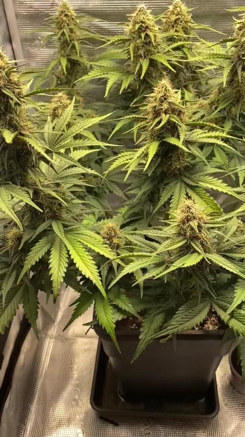
She's looking extremely healthy enjoying lactobacillus made by me and florians living organics full of 10 types of mycorrizae,humic and fulvic ácidas,aminoacids,vegetable hormones and beneficial fungus,also guanokalong extract which is also full of life man! She's just an organic happy girl in a soil full of micro life,thank you everybody peace ✌️ 💚 ❤️ 💛

Processing
Likes
24
Share


@DaddyPrime
Follow
9/2 allowin showing colors finally, getting purple I some fanleaves, she's smelling super sweet like berries
9/4 she's getting so so frosty. Shes growing/budding like a typical sativa. Super Long slender nugs with ridiculous amounts of frost/crystals covering all surrounding sugar leaf. Yet still small and slender actual bud formation. I am very impressed by the resin production so far though. (Sativas always look Impressive(crystals) but lack actual bud structure)
Watered and introduced beastie bloomz to the mix instead of open sesame. Hoping to pack on extra weight.
On a side note: the clone from this plant is flowering as well. I have limited the addition of most nutrients besides what was already in the soil. Its cool to note that the clone that hasnt been fed flowering nutes like the mother plant has, is lacking in smell, color and resin production, in comparison between feeding with open sesame vs not feeding besides water as needed. Its not surprising but i love seeing the proof


Likes
9
Share

@RBG
Follow
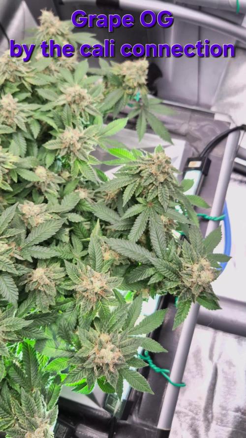
🌿 Grape OG by The Cali Connection 🌿
Week 14
Flower week 8
Flower-day 1
3/12/25
( pisitils showed 25th of October) im timing from pisitils showed but diary runs from 12/12 light flip
No nutrients change today, I'm thinking about 2 weeks or so of feeding, maybe 2 more horti rawk doses before switching out to new millennium winter frost then onto flush with plain water/ph
9/12/25
Dumped nutrients, winterfrost 500ml ( 10ml a gallon) will use for 7 days then flush for 10 or so
Not sure, not sure. Maybe even give LA and Grape an extra week to get Bellini over the end instead of pulling a little early or pulling her as shes finishing up.
Next horti rawk dose probably saturday (48hr run ) then dump then new nutrients.
This is currently week7 day 1 flower from 6/12/25 ( pisitils first shown )
8/12/25
Will be dumping nutrients tomorrow, using winter frost then onto flush


Likes
Comments
Share


@HerbenTrees
Follow
I feel for it being week 5 my builds aren't a fat as I want nite to be I don't no how u guys get suck fat colas can any body tell me wat am I doing wrong because I see some of ur diaries and by week 5 of flower they have massive buds already in a dwc set up

Likes
14
Share


@Bigbombbudz
Follow
Another week closer....
These girls have been a pleasure to grow so far they love everything you can give them ...no deficiencies no problems.....earlier in the week a slight tip burn but after 2 days they adjusted and ask for more.....as they are eating up the nutrients I am replacing and upping it each time...pushing these girls and they are showing there love....17 days since I switched the light and they are filling in nicely.
Until next week, smoke a fatty, help out you're fellow grower

Likes
7
Share


Coming along nicely in a week or so I will Super crop and scrog shortly thereafter flip to 12-12
Supercropped November 9th 2018
Sugar Black Rose clones humidity dome
Lollipoped and Topped and Scrogged
Nov. 11, 2018

Likes
202
Share


@CrazyHorse
Follow
Hello my beautiful friends 😊 😊 😊
2nd week arrived and he is here 💪 💪 💪
Girls looks very happy and I am happy with them 🙂
Day 9- like you can see on picture a humidifier was temporarily removed. I have to clean and maintain it, so humidity is low but tomorrow unit going back inside 😇 😇 😇
Please have a beautiful day and I wish you all to have a fantastic Christmas 🎄 ♥️ 🌲


Likes
16
Share


@Daddyhancocc74
Follow
Day 21 Of flower I did a lollipop / heavy trim (feel like I could have done a lil more) and I have to say I'm running out of room with my lights it's pretty much at 9 inch away from the tallest part of the plant the ppfd high but using a phone app But not seeing no burn on the leafs The intake of water has been up to a gallon an a half (always do some kinda of runoff) pH has been around 6.1
Day 23 Of flower and I added the AC infinity fan (day 1 of fan(actually couple hrs and I think I been missing out on a oscillating fan so get one) still been slowly uping the dimmer it's been getting warmer in the tent so I turned my AC on
NOTES: ( btw it's April and I live in a new apartment and pretty much start growing after a month or so of moving in(moved in November started in the middle of January) and seems like this place holds heat so it's going to be a hot summer) but I can see myself down the road getting a (portable AC for the drying if I want to reach that 60°/60% ) plus that dehumidifier kicks off a some heat and my dumbass is growing in the room I sleep in so since all this I've slept on my couch a few times and plenty more to come

Likes
13
Share


@Malcom
Follow
My first plants! 4 weeks! I love it ! ❤️❤️❤️❤️❤️❤️❤️❤️❤️❤️❤️❤️❤️❤️❤️❤️❤️❤️❤️❤️❤️❤️❤️❤️❤️❤️❤️❤️❤️❤️

Processing
Likes
14
Share


@Nookandplant42o
Follow
previous watering was fed with liquid nutrients, my temperatures are not low so the nutrients come slowly with some intervals giving only water without much runoff occurring a mixture of soil and mycorrhizae for better performance i know i needed calcium and magnesium good the problem was money, i will continue with epsom salt the option i have direct sulfur in the soil has helped me hear a visible change.
water ph 6.2 solution temperature 20 ° - 0.50 g of great white mycorrhiza 2 days earlier were sprayed with water without ppm via leaves - lights off to serve as a cooling shower in the heat is great for stomata .
Simple led panel added as main light source 260watts being divided to 3 plants in the fullspectro tent model, did not get much difference in temperatures compared to HPS, is more the same spectro ratio and UV to reach more directly in the trichomes and terpenes.
Large leaves, like a true sativa even if they are long, go in equally straight and thin shape, trichomes with glands larger than their sister, but this one in front really does not hear physical stress from something just high temperature training, but are very resistant To this, I imagine later on taking this same genetics at favorable temperatures and hydro. A strong smell that I will have to turn to buy a filter, I can not with curious and malicious neighbors need to really contain the smell.


