The Grow Awards 2026 🏆 


Likes
Comments
Share
@Aedaone
Follow
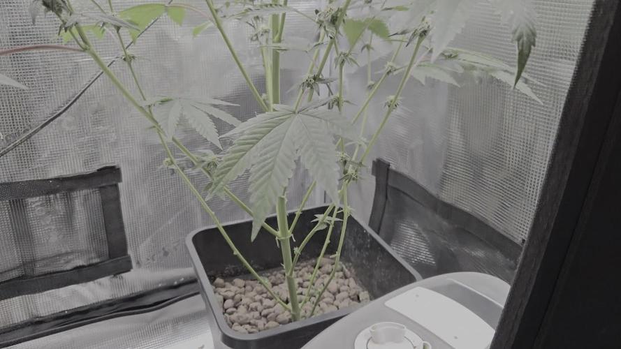
This week wrapped up the generative two phase or midflower and began the generative three phase. The plants were fed the nutrients listed daily. I like to keep the p and k up high during the first week to twelve days of generative three. This week the weather has been more cooperative but far from ideal. The highs have ranged from mid 70's to 90 degrees f. The nights are running low to mid 60's f with humidity in the 90% range at night. Humidity has stayed somewhat high during the day ranging from 70% to mid 50% a few days only. This weather has really been testing these girls resistance. Plants 1 and 2 have been immune to pm. Plant 3 has responded well to treatment with arber. All the plants got a medium defoliation which helped. Everyone stacked this week. They have all reached their maximum height. I used the height of the tallest 87.5" for the official height. The two shorter plants are 61 and 62 inches.

Likes
2
Share
@igrowcan
Follow
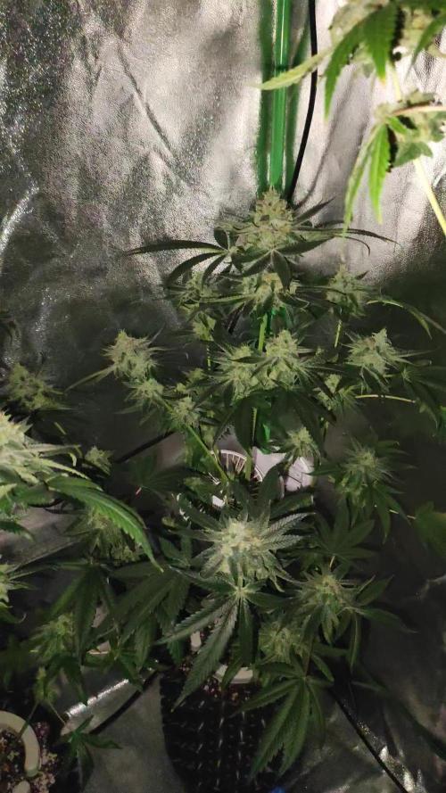
[Week 5 - Rapid Growth]
In just one week, the Fat Banana plant experiences its biggest growth spurt so far within the minigrow box. The
leaves become lusher, and the stems grow stronger as the plant reaches for the light.

Likes
2
Share
@Autower
Follow
Missed a week as been busy but here they are coming into week 10 time to start hitting them with some overdrive for a few weeks before flushing with ph,d water
Likes
29
Share
@DOOBS_N_BREWS
Follow
My overall impressions of this strain from MSNL:
Ive noticed that MSNL genetics tend to switch to flower earlier than all the ther autos i've grown. The initial growth on the plant starts out small going into flowering but then it starts to explode in growth. Because of the fast flip I start my feed at week 3, I used the full NFTG advanced feed, but only at 1/2 the recommended amounts of the early veg schedule they made. This plan of attack worked out well with these plants and by week 5 they could handle the full Nute schedule. A proper flush was needed at-least twice a week during the flowering period. It is a lot of work to maintain PH and soil PPM but its essential with the NFTG and if you do it right you WILL get rewarded with Danky goodness.
I did the same exact training for each of these plants but right off the bat it was clear I had two very different phenos. Plant A grew very compact with extremely tight node spacing and barely getting to a foot in height by flowering. She was kind of tricky to train initially but I was successful at LST and light defoliation and that was pretty much the extent of the training she had. She grew to be short, stout, and plump AF with way frostier trichomes then her sister. Plant B was real easy to LST because she grew with a very wide open body type to begin with, the polar opposite of Plant A to be frank 😂. I was able to shape her easily and It seemed very successful due to the sure size of her buds ! I had close to 10 main colas on her that ranged from 6-10 inches long and she started to tilt over cuz of the weight towards the very end 😅
Overall I would definitely recommend using LST to some extent with these genetics because of the small initial growth you get at first. Start the nutes sooner than later. maybe week 3, but at half the strength. Feeding the soil would be a good idea right from the get go. Mammoth P is a cool microbe that targets phosphorus directly and recharge will slam pack your substrate with beneficial microbes and ive noticed a very clear difference after incorporating these into my NFTG feed. Another thing I did that seemed to help was weekly epsom salt (magnesium sulfate) sprays on top of my bloom khaos foliar sprays. I didn't deal with any deficiencies at all this time around following this feeding regiment 👌I also didn't notice any popcorn nugs on any of my stalks and I contribute that to my feeding schedule and the Spider Farmer SF2000 202W qb , that thing seriously blew these plants up! would recommend 100% !!
Dubes~
*UPDATES*
March 1st- I removed the two GDPS from 32 hours of darkness and chopped branches to hang and dry. Plant B had a wet weight of 19.9 oz! while Plant A finished at 12.1 oz wet(this is with branches and leaves still on obviously). Im attempting hang drying for the first time because my drying conditions will work better with this method. Im using my flower tent with the inline fan/charcoal filter for air circulation. Also gives me the ability to dry in complete darkness.
March 7th-Trimmed both plants today after hanging for 5 days at 40%RH 62-68F. Plant A yielded 1.9 oz (54 grams) dry and Plant B yielded 3.5 oz (98 grams) so a grand total of 152 grams !!
March 14th(1 week of cure/burping)- flavor update 1 week in of curing .. Pheno B is putting out huge diesel and berry aromas almost a little herby but mostly berries/diesel . Flavor is nice with hints of incense , berries, and diesel .. really good joint smoking week. Pheno A is completely different Than B , I do not get any diesel / berry smells at all its more of a fresh pine / herby smell. Im more keen for Pheno Bs smell because its more pungent and stands out a lot more but they both make excellent smokes and get me super stoned!
March 20th(2 weeks of cure) - Yup this stuff is great. I love rolling Js with this shit because it gets me right where I need to be without needing any more. I can't believe how dense I got these nugs to be compared to my last run. I incorporated c02 into this grow half way through so maybe that helped achieve the density but one small nug fills my grinder up with fluffy danky bud!!
Match 31st- (4 weeks of cure) - Both phenotypes ended up fantastically dense. One little nug fills the grinder 100% I would say this is the perfect smoke for anxiety and relation. The green pheno didnt have a berry flavor at first but the berries definitely come out. Both phenos have different aromas but similar tastes for sure. im still undecided on which pheno I like more. The purple one stacked up huge colas and out weighed the green pheno but the terps on the green pheno are way more pronounced and I even think the high is a little more intense.
April 7th- (5 weeks of cure)- The money spot. all this weed is premium quality compared to my friends "street purchase" bud. I am giving this away to multiple friends and I can't get them to stop bothering me for more haha. Definitely a great relaxation aid and sleepy time medicine if that's what you need, seems like most ppl need their night smoke before bed these days so this is the strain they need! 100%

Likes
23
Share
@GrowGuy97
Follow
Wish the buds would have got a little bigger but feel like that’s particularly my fault for a short Veg but overall I am blow away by the outcome for all the seeds to just be random bag seeds! 2 of the plants are drying now 3rd one will be cut tomorrow & the other 2 got probably 2 more weeks! Will do a taste & smoke report and also give a weight as soon as they dry & cure a little bit but honestly couldn’t be happier with my first grow! Thanks for all the support & help along the way & happy growing friends!🤙🏼✌️🏼🌱
Update on 1st plant - dry weight 42g, still smelled pretty Earthy when I put it in the jar but the smell is definitely coming out now, extremely sticky & frosty buds❄️: 1:🏼🙏🏼
Update - plant 2 Dry weight was 40 grams! Extremely happy with the out come this is honestly some of the best bud I have ever smoked! Amazing to me this came from a random bag seed, it has a very citrusy flavor, smells & smokes amazing! Honestly a 10/10 in my book, wish I could knew what strain this was so I could grow it again!😫❄️✌️🏼🌱
Update - plant 3 Dry weight 34g, smells & look phenomenal, the buds are much smaller & had the smallest yield so far but this is by far the best smelling so far! Will have a smoke update on this one soon, stay tuned friends & happy growing!✌️🏼🌱
Update - plant 5 dry weight was 52g it dried a little faster than plant 4 which I will probably jar tomorrow! Will update again with a smoke report soon, this plant brought my total so far up to 168 off 4 plants👍🏼 Thanks for following friends & happy growing! Also the butter I made from the trim was 🔥🔥
Likes
7
Share
@TiRobotProds
Follow
20/11 Ce petit se développe super vite, irrigation a l'eau uniquement tous les 2/3jours.
Bientôt transplantation pots 6L pour plus de développement. Reste sur le réseau growers.
26/11 repiquer dans un pots de 6L dans du ATAMI - Janeco-Light Mix qui es super pour développer davantage de racine . Arrosage a l'eau ph 6,5.


Processing
Likes
16
Share
@CzAlmighty
Follow
Tak jsem se dočkal. Bez žádných velkých problému jsem prošel 5tydnem . Už lehce voní 🍀 moje návraty z práce jsou vždy luxusní 😀❤️Jinak krasne reagují na LST. 👌🏼


Likes
12
Share
@Luke_Lee
Follow
-15.08.2024
The light intensity has been reduced so that the ppfd value is below 1000.
The soil still felt moist, the plants were not watered.
Yellow leaf tips are still there, even turning white.
Continue to observe, do not fertilize and research.
-16.08.2024
Did nothing
-17.08.2024
Visual Control
Soil was dry, Both plants get watered with 500ml water per plant (no nutriens)
-18.08.2024 - 20.08.2024
Did nothing, just Visual Control
-21.08.2024
Soil was dry, Both plants got 1Liter Water per Plant.
(1ml BioGrow, 2ml BioBloom, 2ml TopMax - 1L Water)


Likes
28
Share
@GrowingisFunkO
Follow
Overall, the garden is doing great. Had a scare with the WW with letting it dry out a little but it looks like its going to be fine.
The pistils are starting to change color and leaves are starting to lose color but the buds are fattening up. I can't wait to see how they all turn out.

Likes
38
Share
@J_diaz420
Follow
2 días entre servilletas y al sustrato.
Aprox 12 hrs en obscuridad y se comienza el ciclo lumínico 👌👊👨🌾🏻

Likes
14
Share
@GoldenWeedGrower
Follow
D50/F06 - 20/05/23 - Nothing to report
D51/F07 - 21/05/23 - Added water and nutes EC=1.1 pH=5.7
D52/F08 - 22/05/23 - Nothing to report
D53/F09 - 23/05/23 - Added water. EC=1.1 pH=6.4
D54/F10 - 24/05/23 - Added water and nutes EC=1.0 pH=6.3
D55/F11 - 25/05/23 - Start the week out. I set up a system to feeding Nora during my stay out (I'll take some picture about...)
D56/F12 - 26/05/23 - I leave today, 1 week away
Likes
6
Share
@HomeGrowHeaven
Follow
The plant has recovered well from the last capping and defoliation🏻♀️
A few leaves have yellowed a little, but that's not so bad🏻♂️
It will get a compost tea🍵 in a few days when it is sent into flower and then everything will be fine again😊💪🏻🏻
The compost tea is brewed and was given to the Watermelon Runtz at day 49 of Veg🍉🏃♂️

Likes
11
Share
@MR_FASTBUD
Follow
This is plant one and two, plant one cane down 5 days before plant two so didnt get the exact wet weight, 5 days into hang dry plant one weighed 250g, plant two was cut down and chopped for box dry, weighed 200g for the heads and 300g for the lower plant produce, will update when dry and tested

Likes
13
Share
@ThCvibez804
Follow
Yo today me and my lady haf a Smoke Session Today And The Vibez Was Great Check The Video


Likes
27
Share
@adam_pawloski87
Follow
Over all was an amazing grow, super sticky dense nugs with smell of berries, highly recommended for everyone!!
Likes
33
Share
@BADKUSS
Follow
Hello everyone,
the plants are still very good, no deficiencies in sight and canopee rather homogeneous.
the stretch is soon over and I'm going to be able to stop the growing fertilizer, for which is knitting, I finished and let nature now play ...
Now place to photos ...
Likes
23
Share
@cadur
Follow
Looks like growth has stopped and the work is going into the flowers.
Some leaf tip burn but in not going to change anything, I could fuss and look at the TDS, but I've always got myself in a tis and mucked things up.
I'd say this is looking to be an average yield. Not the smallest, not the largest!