
Likes
Comments
Share


@Ambz_1990
Follow
Just approaches week 7, I don't know when to initiate the dragon force, kill feed and then flush, iv gotta buy a camera lens this week to check trichomes but I'm nearing the end

Likes
1
Share


@fedee420
Follow
ESTA SEMANA SE HACE TRASPLANTE A 10 LITROS Y PASAN A LA SALA DE FLORACION
Likes
24
Share


@DogDoctorOfficial
Follow
🌱 Divine Seeds Week One Report 🌱
Hey, fellow growers! 🌿✨
We’re back with the Week One update on our exciting journey with Divine Seeds! Our Moon Rock, Opium, Fractal, and Big Demon seedlings have made remarkable progress, and we’re thrilled to share their growth story with you.
🌟 Week One Highlights 🌟
This week has been all about establishing a strong foundation. With the help of the amazing CannaKan method, our seedlings are thriving! Here’s a closer look at their journey:
1. Moon Rock 🚀
• Growth: These seedlings are reaching for the stars, displaying vigorous vertical growth.
• Health: The leaves are vibrant and green, indicating excellent health.
• Notes: Moon Rock is showing great potential for strong stem development.
2. Opium 🌸
• Growth: Opium seedlings are flourishing with grace, showcasing steady and balanced growth.
• Health: The foliage is lush and healthy, with no signs of stress.
• Notes: Their elegant growth pattern is a joy to watch, promising beautiful plants ahead.
3. Fractal 🌀
• Growth: Our shy Fractal is slowly but surely making progress, showing unique leaf formations.
• Health: Despite a slower start, the seedlings are healthy and adapting well.
• Notes: Fractal’s unique patterns are starting to emerge, hinting at its distinct genetic traits.
4. Big Demon 💪
• Growth: True to its name, Big Demon is growing strong and bold, with robust stems and leaves.
• Health: The seedlings are exceptionally healthy, with deep green leaves.
• Notes: Big Demon’s rapid growth is impressive, setting the stage for a powerhouse plant.
🌟 Key Observations 🌟
• Environment: Maintaining optimal temperature and humidity has been crucial. The CannaKan method’s stable conditions are proving to be a game-changer.
• Watering: Careful watering practices are ensuring the seedlings receive the right amount of moisture without any risk of overwatering.
• Light: Providing adequate light has been essential. We’re using a balanced light schedule to support healthy photosynthesis.
🌱 Next Steps 🌱
As we move into the second week, our focus will be on:
• Nutrient Introduction: Gradually introducing nutrients to support further growth.
• Transplanting Plans: Preparing the seedlings for their transition to larger pots.
• Continuous Monitoring: Keeping a close eye on their progress to address any needs promptly.
Stay tuned for more updates as our Divine Seeds continue to flourish. We’re excited to share every milestone with you on this incredible journey!
Happy growing, and may your gardens be ever green! 🌿💚
#DivineSeeds #WeekOneReport #CannaKanMethod #MoonRock #Opium #Fractal #BigDemon #GrowDiaries #PlantMagic #GreenThumb
Germination method 🌱 @thecannakan
Genetics
@divine.seeds
Nutrition
@aptusholland 🌿
@aptus_world 🌎
@aptus_es 🌍
@aptusbrasil 🌱
@aptus_thailand 🌿
@aptus_portugal 🌳
@aptususa_officiala 🍀
@aptusplanttechnz 🌺
@aptusplanttechaus 🍃
Ambient controls🎮
@trolmaster.eu @trolmaster.eu.support @trolmaster.support @trolmaster.agro
Soil @promix_growers_eur @promix_cannabis
LED - @lumatekeu
Watering- @autopot_usa @autopot_global
Love and attention- @dogdoctorofficial
#aptus #aptusplanttech #aptusgang #aptusfamily #aptustrueplantscience #inbalancewithnature #trolmaster #trolmastereurope #trolmastersecrets #Autopots
#GreenJoy
As always thank you all for stopping by, for the love and for it all , this journey of mine wold just not be the same without you guys, the love and support is very much appreciated and i fell honored and so joyful with you all in my life 🙏
With true love comes happiness 💚🙏 Always believe in your self and always do things expecting nothing and with an open heart , be a giver and the universe will give back to you in ways you could not even imagine so 💚
More info and complete updates from all my adventures can be found ⬆️link in the profile description ⬆️
Friendly reminder all you see here is pure research and for educational purposes only
💚Growers Love To you All 💚

Likes
393
Share


@FoTwenny
Follow
I apologize for the video quality. I have been super busy, but still wanted to make something nice for y'all!
Day 56: Gave the girls another plain watering pH adjusted to 6.5 then added Mammoth P @ 67°F. Not fading as quickly as I'd thought they would.
Chopped the Triple Cheese today and moved our Gelat.O.G. into the open spot in flower. Breeder specs show 50-56 day Bloom an I think last run she may have went too long.
Plan to be in the garden 8/5 (Day 58) and hoping to see some more fading and swelling going on. The aromas on the tent are absolutely tantalizing. I will try to get a few more photos for ya'll.
Day 58: Did some minor defoliation in the veg tent and took photos. I decided today I would upload unfiltered and unedited photos. Enjoy!
Day 61 of 12/12: I noticed seeds in the Skunk #1.😭😭😭 Possibly other plants too, but I was mostly on the lookout for the flowers to tonight. I noticed a single banana popping out of Purple Queen. I decided to harvest all but the Strawberry Amnesia and the Blueberry. I will get pictures soon and update with harvest deets.
I need to take a closer look to see what plants may have been pollenated and if there are any other nanners. I did have a timer fail (which has been replaced) at the end if week 1 of flower. Not sure if this could be the cause, if I let her go too long, or if it's just the genetics. I am hoping for minimal damage. I'd be interested in a Purple Queen x Chenobyl cross. Think the seeds would be prone to hermies or would ya'll toss the beans in the bin?


Likes
22
Share


@dataTwiiix
Follow
06/27: the small buds dry up quickly with the current climate near my house.... So I was able to taste the famous apple fritter. And I'm glad I dont know when I grow another AF. because what I like is to taste the novelties

Likes
26
Share


@OGbros
Follow
The plant looks good. In these days the temperature outside are very cold (about 5 celsius), but in the box there is a electric radiator connected to a control unit (inkbird): every time the temperature goes under 20.7 C the radiator turn on. Yesterday I broke the holding pulley of the lamp😰 so I had to hold it with a wire. it will remain that way for 2 days, I've already bought a new pulley from internet. Anyway I defoliated the purple punch to bring the light to the apicals. Every 3 days it puts out new leaf, incredible! I used 10ml of flora grow , 6ml of flora micro (hard water) 5ml of flora bloom and 5 ml of cal-mag directly in the solution
edit: I resolved the problem with the lamp. I top up the solution with 10l of water in wich I put 20ml of flora grow, 12ml of flora micro, 6ml of flora bloom and 10ml of cal-mag. I also used 10ml of pH- directly in the solution to lower the level of the pH (6.3-6.4 at the moment). Everything look ok😀


Likes
4
Share


@CrazyDutti
Follow
0.25g Bittersalz zusätzlich .
Habe die letzten 2 Wochen , Probleme ( siehe Bilder ) . Bin noch am lernen wie ich der Pflanze helfen kann. Hoffe auf Besserung - Finger kreuzen 🤞🏻
Likes
5
Share


@Ozgrow
Follow
Filling out need a second screen. Dont think will have too much more stretch. The white choco haze is still behind no way mear as much foliage, however considering her start to life she is doing much better. Gorilla looking nice and the 2 choc mints of the left of the photos are looking terrific.

Processing
Likes
44
Share


@I_and_I
Follow
First grow completed, turned out far better than I could ever have hoped, the plants totally bossed it for all the winging it and improvising I put them through, but we got there in the end haha,
Looking forward to next grow, can't wait to enjoy using all good genetics, already have some Bruce banner #3 seeds ready to roll out :)
Thanks for reading
Likes
17
Share


@HowtoBubatz
Follow
Week 6 of flowering, and things are heating up—literally with the scent! 🌬️💨
Both plants have significantly increased their aroma, and every time I open the grow box, I’m greeted with a strong and delightful smell that fills the space.
The non-LST plant is much further along in its development. Its buds have progressed beautifully in color, and I’ve already noticed the first amber trichomes appearing. 🍂✨
This, along with its overall appearance, is signaling that it’s nearing maturity. Because of this, I’ve started flushing the plant with clear water to prepare it for harvest.
Speaking of the LST plant, it’s about a week behind. 🌱
While it’s not as far along, it’s still thriving with strong trichome production and a stunning Lemon Haze aroma that’s sharper every day. 🍋💚
Both plants are doing great, and the differences in their timelines make this grow even more interesting. I’m excited to see how the LST variant catches up in the coming weeks! 😊🌟

Processing
Likes
11
Share


@Newbie5648
Follow
Day 57. Nutes increased for more growth hopefully.
Day 58 notice alot what looks like thc from leaves and hairs.
Day 59 to 63 see more orange hairs and trychomes. See videos
Average 200ml water everyday.


Likes
8
Share


@Kountryboi90
Follow
Day 35 and 12 days into flower and the girls are looking fantastic. One turned out to be a photo so I've removed it and put in my other tent. The are seemingly loving the Advanced Nutrients. I've fed twice at full strength and I'm nothing growth every day. At about day 21bof flower, I will do another and last defoliation of these girls.


Likes
6
Share

@MindFlowers68
Follow
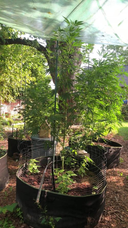
The honeydew fallout from the spotted lanternflys in the maple tree above have gotten out of hand. It's almost a constant mist. The only thing I could think to do beside moving them like an ancient Egyptian was adding a protective canopy of some sort. I wanted to get the clear poly weave but this green one was half price so went with it instead. With the way I have it oriented, it doesn't really block out that much direct sun too. In the morning and afternoon the angle of the sun is able to shine right on the plants. It's kind of a win win because should decrease my chances of mold later down the road towards harvest time if it does decide to rain a bunch. I tried washing the plants once this week with some light soap and water with a little baking soda mixed in. I rinsed with cone setting with hose right after and the plants were praying next day but i didn't have canopy up yet so they got hit again. I spotted several pale green assassin bugs on my plants so now Im a little nervous about washing the plants again because I want to keep them around. They are nymphs right now I may try to collect the ones i see become cleaning the plants one last time.
Hanging that canopy was one of the more frustrating things I've done in a while. I wasn't sure If or how it would work but I has some medium gauge synthetic rope I tied to the eyelets and eventual 4 tree limbs. I had to bounce back and forth to get it tension climbing my fruit trees and 8 foot ladder. I originally disassembled an unused tomato trellis i built to use some of the lumber to build a post to tie off on in one corner but it ended up being at the wrong angle relative to the tarp and I started to get worried if a storm ripped it out of the ground. So I opted to used only the surrounding trees. After getting it all tension I realized that it was to far to one side leaving two of the plants almost exposed, so I had to come back the next day and re adjust but now I have it in a pretty good spot. I survived a decent storm already this week. Just hope it lasts. Overall making outside feel slightly leas stressful and anticipating more of a good harvest rather than just an experiment with my first outdoor grown.


Likes
47
Share


@Master_weeda
Follow
Bonjour à tous les padawans et maîtres jedis
Super plante ou plutôt jolie petit arbre avec de branches bien chargées de grosses têtes remplis de trichomes
Un vrai plaisir à cultiver très belle et résistante
Likes
12
Share


@Jays_Not_Here_Man
Follow
Week 7 of Veg for these beautiful ladies! It was a stressful week for the girls but they made it❤️
Day 44 I decided the girls had outgrown their 2 gallon nursery pots and were ready to move on. After a quick cleanup of the undercarriage, they were ready to go.
I like to keep my soil in a Rubbermaid bin, either with a fan lightly blowing, or the exhaust line from my tent blowing at it. Since I started doing this, I have not had any gnats or pests in my grows, of course with regular cleanings and IPM…
This time around, I decided to use some Dynomyco to increase the mycorrhiza count of my Promix HP. This will increase root development and allow for better uptake of nutrients overall. Within an hour or so all of the ladies were transplanted ❤️ 2 in 7 gallon fabric pots, and 1 in a 5 gallon fabric pot ( the 5 gallon will finish in my 2x4 tent)
Once the girls bounce back and take hold of their new forever pot, I will be setting up my custom “floating” SCROG net and allowing them some time to get bent and tucked all nice snug before I flip to a flower schedule. Happy Gardening ❤️🇨🇦🌱😎

Likes
9
Share


@TiRobotProds
Follow
4ème semaine de floraison. Cette dame ce porte a merveille ☠️☠️💀 irrigation tous les 3 jours environ 6L, 12L/Semaine.

Likes
49
Share


@masterofsmeagol
Follow
7/11 Got half in of rain last night. Glad I didn't water. Ph of rain water is very acidic. Added supports to the blueberry cheese in the 50. I just used string and went diagonal and attached to the cage. Wrote a ton but it disappeared. Anyway looking at previous diaries I was wrong about senescence. It wad the life cycle of those earwigs that did that to my plants (see other diaries). This soil mix is amazing. These plants gave only gotten like two small feeds of big bloom. I showed my commercial buddy and I'll keep it between us what was said but it made me feel really good. I'm considering either expanding the cage in one direction with some lumber I already have or moving a couple outside tge cage. That way I have more room. He said he's seen plants structurally similar reach huge heights (14f) so I'm just trying to avoid future problems. I at least need to get the trellis on. I hope poor that's food enough. Did a real nice video this morning but nothing wanted to upload and it just uploaded the same one twice. I'll try again and hopfully it will go up. If not I'll put it up tomorrow.
7/12 Dad has surgery this morning. I did a quick video and took some pictures. I need to do some rearranging with the plants and cut a pallet to fit in the back. Then I can put a vertical trellis up. Haven't decided if I'll add on to the cage, remove a couple plants or just rearrange things but I'm leaning towards the later. They will need water again and I'm wondering if I should start nutrients but everything looks so good. By this time on past diaries I'd be losing all my leaves bu now. MI think it wad part ear wigs and part to many nutes. This soil mix is taking these plants through veg. I'll probably do a feeding soon but it will be organic and it will he small like a big bloom or ancient amber. Im leaning towards not using growbig this year as my plants seem to be doing great without it. We'll see. I'll keep this updated.
UPDATE: Went back over and gave plants a full watering. Some were slightly drooping. I was going to add nutes but decided against it as I didn't see any deficiency. So far NO Earwigs! I comed through the plants and I did find a jpn beetle which I happily killed. I mixed up 8 gallons of water and gave it to the 11 plants so it wasn't quite a gallon a plant. I need to rearrange the plants so I have room to move around. I also need to cut another pallet and use the spaces I'm not. Trellis needs to go up.
7/13 I think I've got the watering amount down. Now just to find how often which will depend on weather. I've watered very little this year. They loved that gallon. I was going to use big bloom and kelp me/you but looking at my garden I decided not too. My buddy asked what i was addingvthings for and to wait fir what i added to do what I wanted it to do. I see no nutrient deficiencies so why add anything? I think this soil mixture will get me all tgexway through veg. I dont plan on using much in fliwer either. Definitely good genetics. I really need to cut that palley and move the 1 10gallon to the far back coener. It will open thibgs up so much better. The garden looked so beautiful this morning. Getting very aromatic. I started untangling trellis netting but had to leave. I'll update what I do. Happy growing folks
UPDATE: Went back over as I had a slight intuition that I needed to check the garden. I found and killed at least 7 jpn beetles. That's what's been making holes. They tried to escape and bounced off a tarp but I got it. I went through the interiors and found two pillars. If these beetles are gonna be around I'm ordering a net. I'm also close to positive I'm going to extend my cage in the back a few feet. Things are getting unmanageable in here. My buddy said I'll be having problems soon if I don't do something. I'll document what I decide to do.
7/14 Found ONE jpn beetle in the garden this morning. I can manage those well enough. The birds help too. I check my garden multiple times a day so I manually remove many pests. However I've noticed some thrip damage on a leaf and a leafhopper damage on "A" leaf so there are "some" pests around but not enough to spray shit. A couple wasps were doing there job while I was there. Supposed to get thunder storms after three and tonight. Supposed to get over a half inch of rain tonight. I lifted the bags and decided to hold off and let mother nature take care of it as the bags weren't totally dry. Only problem I really have is space. I AM moving that 10gallon (that's the same size as some 20's) in the back. There's 27in not being used and a few feet the other way. My buddy cautioned me that I'm going to have problems since my plants are so crowded. I agree with him. I spoke with me father and we have most materials to extend my cage four feet in the back. I think that's my plan. I'll extend the structure before the stretch then I can put up the supports. We'll see how this goes.
UPDATE: Went back over to check the girls as I had a feeling I ought to. When I got there I saw that a couple of the blueberry cheese were pretty light (liftng the smart pot) but the others seemed to be fine. ONE 10th planet was light like that and the purple punch in the 10 gallon was as well. Each plant thar needed it got at least a half gallon of water. I'm waiting to see if we get the thunderstorms and the half inch of rain. I watered the MASSIVE blueberry cheese in the 50 but I only gave it 1 pitcher which is like 1/4 gallon or so. Don't know why I even gave it that. Looked fine but the soil WAS pretty dry. Next year I'm giving myself way more room. I was running trying to chase these jpn beetles. This time I have the dawn and water and a measuring cup to knock them in. This ain't my first rodeo. I did notice some bright yellow streaks on a leaf edge and I'm hoping it's not septoria. I doubt it but I have an anxiety disorder and I worry. I hope I can get the cage extended sooner rather than later. It's getting hard to move in there and more importantly I can't take any more plant pathogen problems. I'm considering starting a plant doctor regimen just to be safe.
7/15 Got a bunch of rain last night. No jpn beetles in the garden and not really much damage. I did notice this (I think it's leaf hoppers) that leave those dots close together on a leaf so it appears I've got a variety of pests. I'm considering how to approach this. I mean the damage is very minimal but I don't want it to get out of hand. Another thing I need to look out for is leaf septoria or any other fungal pathogens. I believe that has been part of my problems in the past. I think that's what caused my earlier grows to drop all their leaves so quick. I think I'm being overly cautious but its very crowded in there. With my father just getting out of surgery the girls will need to wait a little bit before before I can extend the cage. I could still cut the pallet and move the 1 10gallon and that would give more room. I rearranged a LITTLE BIT so they have a little more room but I've really got to get this cage extended.
UPDATE: IT Rained so hard I had to pull the car over. We hydroplanned the whole way home. After working ob my house I went to see what the damage would be like. NOT A SINGLE BREAK THAT I COULD SEE. I took a video but since the wifi here sucks I'll have to upload it tomorrow. Walking around in the cage even if I cant get it extended I think I'll be OK. It obviously will open me up to lots more issues but at the very least I can reorganize before I build on. The 10 gal purple punch would fit perfectly in the back and I have a pallet I can cut to fit it in place. Putting that one back and pulling the others forward will be much better than what I've got going on now. I'm also thinking about running an extension cord and putting fans under the canopy or at an angle to keep the wind moving. Just thinking outloud. However after that storm the girls looked as happy as I've ever seen them. All happy praying to the sun, thankful for the much needed rain. Mother nature does a pretty good job with out me messing with it. I've noticed a couple interior lowest leaves turn yellow and die like a nitrogen deficiency but everything else is fine. Also noticed a leaf that looked like a p deficiency but again, it was the VERY bottom leaf on ONE plant. Again the rest of everything looks fantastic. I'll keep an eye out for anymore nutrient deficiencies and if anyone that reads this sees some please let me know. I should've taken stills since they looked so good but I got it on video.
7/16 It POURED last night and throughout the day. TORRENTIAL rain. The branch breaking sheet rain that us outdoor growers learned to fear. My plants aren't trellised currently. I know what I need to do now. I have a pallet to put in the back corner and I'm moving the purple punch in the 10 there. And pulling others forward where there's more room. Then when I extend it (it's gotta be done this week) everything g will be in their proper place and I can just throw on a vertical trellis. I also noticed more (leafhopper) damage on a leaf. Different leaf of the same plant so I'm considering spraying something. I have a number of products but I was trying not to use them. Luckily I have these diaries so I can look back and see certain plants reactions to certain nutes or fungacide/insecticide/nutes and the doses used. I haven't been using much but if my plants will remain cramped I'm going to start the plant doctor. I'm seeing more pillar damage too but BT is super narrow so I'm thinking when I fo spray for pests I might use cap jack and be done with it. Then I can apply the BT in flower if it's necessary. I took a video but I have to wait until tomorrow to upload. I took a quick snapshot though.
7/17 Despite the torrential rain I don't have any breaks. I'm noticing more pest damage though. Another leaf on the same plant had those closely shaped round circles. I forget what pest it is but it's there. Caterpillars are there I'm sure so I may do a preclcentative spray. Just unsure what I'm going to use. I lost a COUPLE very bottom interior leaves that look like they just got used up. After this rain I think think the plants might benefit from a feeding. Probably next water after they dry out. I REALLY need that cage extended. I expressed that today and it should be done this week. I'm looking for pallets today. I have the little one that I can put in the back which will allow me to move the 10 gallon and move the other forward. That will help some but I need more room. I'll update as I go.
UPDATE: GOT A SMALL PALLET AND IT FIT PERFECT IN THE BACK ROW. I MOVED THE PURPLE PUNCH IN THE 10 GALLON ONTO IT. I SHIFTED A BUNCHVIF THINGS AROUND. I ROTATED ON BLUEBERRY CHEESE 180 DEGREES SO IT WOULD FIR BETTER. CROP ROTATION IS GOOD ANYWAY. I TOOK VIDEOS AND YOU CAN NOW SEE THE ROWS MUCH BETTER. 2 WITH 3 and 1 WITH 4. IT'S SIGNIFICANTLY BETTER THAN IT WAS. I CAN GET AROUND ALL SIDES OF EVERY PLANT NOW. GRANTED THE LARGE 50 IN THE BACK IS GOING TO HAVE SOME TROUBLE BUT ILL JUST STAKE IT TO THE CAGE. IT WILL GROW TOWARDS THE SUN ANYWAY. IM SUPRISED I DIDN'T LOSE A BUNCH OF LEAVES AFTER THIS RAIN. MOVING THINGS AROUND AND LOOKING ON THE INTERIOR OF PLANTS I FOUND A COUPLE LEAVES THAT HAD BEEN USED UP. I REMOVED A COUPKE LEAVES THAT HAD DONE THEIR JOB. I'LL UPDATE AS I GO ALONG.
