
Likes
Comments
Share


@MadeInGermany
Follow
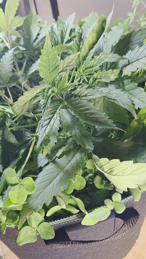
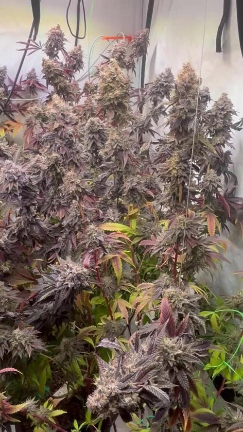
Flowering day 52
since time change
to 12 / 12 h
Hey guys :-)
All ladies look very healthy and delicious :-)
We're slowly getting closer to the end.
This week it was poured 3 times with 1.2 l each (sewing materials see table above)
At the next watering there is a very light rinse with Clean Fruits so that the last nutrients can be slowly used up.
The trichomes are now checked every day.
I'm looking for my microscope with an app so I can take pictures of it 👍.
I wish you a lot of fun with the update and stay healthy 💚🙏🏻
👇🏼👇🏼👇🏼👇🏼👇🏼👇🏼👇🏼👇🏼👇🏼👇🏼👇🏼👇🏼
You can buy this Nutrients at :
https://greenbuzzliquids.com/en/shop/
With the discount code: Made_in_Germany you get a discount of 15% on all products from an order value of 100 euros.
👇🏼👇🏼👇🏼👇🏼👇🏼👇🏼👇🏼👇🏼👇🏼👇🏼👇🏼👇🏼
You can buy this strain at :
https://www.exoticseed.eu/de/hanfsamen/hippie-therapy-cbd
Water 💧 💧💧
Osmosis water mixed with normal water (24 hours stale that the chlorine evaporates) to 0.2 EC. Add Cal / Mag to 0.4 Ec Ph with Organic Ph - to 5.8 - 6.5
MadeInGermany


Likes
8
Share

@420DeepGrow
Follow
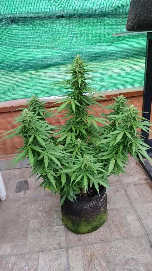
📆 Semana 5
La fase de engorde se hace evidente y los cogollos aumentan de tamaño de forma visible, uniéndose progresivamente a lo largo de las ramas. La producción de tricomas se intensifica y los aromas comienzan a ganar mucha presencia, mientras los pistilos siguen apareciendo en abundancia.
La planta centra prácticamente toda su energía en el desarrollo de las flores, manteniendo un elevado consumo de agua y nutrientes. Una nutrición equilibrada y un buen manejo del riego serán fundamentales para aprovechar al máximo esta etapa de máxima demanda.
⚡ EC: 1.7–1.9
💧 pH: 6.2–6.5
🌡️ Agua: 18–22°C
🌫️ Humedad: 40–50%
☀️ Luz: Fotoperiodo natural, abundante sol directo
🔥 Nota: El ritmo de engorde continúa acelerándose y la producción de resina aumenta semana a semana. Mantener unas condiciones estables permitirá que los cogollos sigan ganando densidad, aroma y peso.
Seguimos creciendo fuerte 💪!
Likes
27
Share


@TheCannaProphet
Follow
------------------------------------
~SEEDSMAN PEYOTE ZKITTLEZ~
------------------------------------
Description
Peyote Zkittlez is an Indica-dominant hybrid strain bred by pollinating Peyote WiFi with the sweet-tasting Zkittlez. Peyote WiFi is itself a cross of Peyote Purple and the US Indica WiFi strain. Zkittlez is a 3-way cross of Grape Ape, Grapefruit and a third, top secret, strain.
Peyote WiFi is an Indica-dominant strain which is a cross of Peyote Purple, derived from a single purple phenotype which was back-crossed for several generations to cement its qualities, and WiFi, a US Indica strain created by crossing The White with Fire OG. This plant is medium-sized with large, broad leaves and takes around 75 days of flowering before it is ready to harvest producing very generous yields of highly potent weed - up to 650 gr/m2 indoors and 1500 gr/plant when cultivated outdoors. The THC potential of this strain is very high with plants attaining 26 - 28%.
Zkittlez is a sweet-tasting Indica-dominant (80%) cannabis strain which produces high yields boasting high levels of THC. This is a very colorful strain displaying green and purple colors with bright orange pistils.
Zkittlez is a 3-way cross between Granddaddy Purple, Grapefruit and Afghani. It is suitable for growing in all environments and takes between 60 - 65 days to complete flowering indoors. Outdoors it will be ready to harvest during late September in northern latitudes. Yields are very good and vary between 450 - 600 gr/m2.
Zkittlez' scent is very sweet and entices with ripe tropical fruits and candy which also follow through in the taste as well. THC production is in excess of 20%. The effect is very uplifting, especially for an Indica strain. Therapeutically it is used to combat stress, depression and anxiety as well as for those with ADHD.
Peyote Zkittlez will make a superb, highly potent and high-yielding Indica-dominant addition to all seed collections which is certain to become a growers' favorite. Get yours from Seedsman now.
*description credit to Seedsman Seeds
__________________________________________________________________________________________________________________________________________________________________________________________________________________________________________________________________________________________________________________________________________________________________________________________________________________________________________________
BREEDER/BRAND: Seedsman
PRODUCT TYPE: Seeds THC
GENETICS: Peyote WiFi (The White x Fire OG) x Zkittlez
VARIETY: Mostly Indica
FLOWERING TYPE: Photoperiod
SEX: Feminized
THC CONTENT: 20%+
YIELD: Indoors: up to 650 gr/m2; Outdoors: up to 1500 gr/plant
GROWS: Indoors, Outdoors
FLOWERING TIME: 60 days
MEDICAL CONDITIONS: Anxiety, Depression, Stress
MEDICINAL PROPERTIES: Yes
EFFECT: Powerful, Relaxing
__________________________________________________________________________________________________________________________________________________________________________________________________________________________________________________________________________________________________________________________________________________________________________________________________________________________________________________
THE SETUP:
~Planted into Jiffy Peat Pellets that were hydrated with de-chlorinated water with SuperThrive added then ph'd to 6.0 @ 80℉
~Grown 100% organic in 10g fabric pots with Mother Earth 70/30 Coco/Perlite medium amended with 2tbs/g of Down To Earth 4-4-4 / 2 cups/g of Earthworm Castings / 1tbs/g of Dr. Earth Flower Girl 3-9-4, 1tbs/g of Dr. Earth Bat Guano, 3/4 cup of Down To Earth Azomite and 1 tsp/g Down To Earth Fish Bone Meal.
~24hr light cycle during Germination / 19/5 light cycle for Vegetation and 12/12 for Flower
~Straight water ph'd @ 6.2-6.8 when needed and weekly Compost Tea's.
__________________________________________________________________________________________________________________________________________________________________________________________________________________________________________________________________________________________________________________________________________________________________________________________________________________________________________________
WEEKLY UPDATES:
9/20- 💥BOOM!💥 Week Six of flower is here and my girl's in high gear, stacking her flowers and pumping out trichomes!
Today I watered her with 1.5g de-chlorinated water with 5ml/g of Botanicare CalMag+ added, then ph'd to 6.2 @ 72℉. I turned her pot and plucked a couple of yellow shade leaves...the basic daily maintenance.
9/22- I didn't water her yesterday as she looked great and was 'praying' hard. Today she was given 1.5g of de-chlorinated water which was ph'd to 6.2 @ 72℉ which I gave her through her drip pan (bottom chuggin) and I also gave her pot a turn.
She continues to pile on the trichomes and is taking on a exquisite purple hue to her flowers! She is definitely one of the most photogenic cultivar's I've grown, especially with her 'Triple Headed flower', and is looking lovelier by the day! 😍
9/24- We're getting close to wrapping up Week Six of flower in a couple of days, which will put her around two or three more weeks from finishing, with the most exciting weeks yet to come!
I didn't water yesterday and today I went ahead and Top Dressed her with 2 tbsp/g Dr. Earth Flower Girl 3-9-4, 1 tbsp/g Dr. Earth Gold Premium 4-4-4, 1/2 cup Down To Earth Bio-Fish, 1/3 cup Down To Earth High Phosphorus Bat Guano and 2 cups of Worm Castings.
I watered in the Top Dress with 1.5g of de-chlorinated water which was ph'd to 6.2 @ 72℉ and let her enjoy her meal! 😜
9/26- After her heavy watering on the 24th I didn't water yesterday and when I checked her today at 'lights on' and she still had some weight to her pot and her leaves were praying hard so I held off on watering today and will hit her tomorrow with her usual 1.5g watering.
~Thanks for stopping in! Things should be getting a lot more interesting in the coming weeks...Stay lifted and be Blessed! 😎🙏~

Likes
6
Share


@4F1M6
Follow
This lady continues to hulk out. As she gained 8 inches extending her bulky frame. Super strong and hardy genetics that's for sure. Those leaves fam!!! Like baseball gloves its just amazing. I had to super crop some of her side branches. Plus her main for the third time SMH. Nothing like stressing the bitch out eh... This time will be the last as I throew on a tie to lock it in place. Should've been done from the beginning.
Shes just starting the fourth week of flowering and she's already well developed. Even the fan leaves are getting sugar coated. As trichome production starts to rev up. Bud sites are beefing up. I already know I'm in for some cannon buds. Muhahahaha.
Treated her with Dr zhymes as a preventative. I also upped her pk intake. Supply this lady a little something extra as she goes off with the flowers. Very happy and healthy lady. Until next update. Happy growing and stay lit fam.

Likes
33
Share


@GanjamanAndWeedmaster
Follow
The second week of the flowering phase is over. I made you a video where you can already see small flowers. it is straining more and more for weed and the plants are getting bigger and bigger.

Likes
57
Share


@Regenwurm
Follow
Nichts besonderes! Die Seeds sind gekeimt wie geplant, wurden dann sofort in Steinwolle gesetzt. Nach 3 Tagen in der Steinwolle waren alle Ladies am Tageslicht. Nach nur 2 Tagen in der Steinwolle direkt in coco töpfe 1 l eingesetzt. Klima ist nicht ganz perfekt, bekomme es aber nicht besser für diese Phase hin. Ventilator läuft 24/7, besonders für die kritische Zeit ohne Wärme der HID Lampe arbeite ich in den Wochen 1-5 mit CO2 Bags, da ich wegen dem Klima (draußen 0 Grad) keine direkte Zuluft nutzen kann.


Likes
21
Share


@Gardener_of_Goodness
Follow
The Fade, the colours, the smells, the resin! What a show! 😍
I hope your enjoying the pics as much as I am, it’s hard to describe the smell now, it’s varnish chemical but with a very strong berry tone. Last week of flush now! Diary update a little late sorry!
Will report next week with harvest after 48 hours in the dark. I’ve watered (PH 6.1) her for the last time today so will let her dry out for a couple of days before lights off.

Likes
1
Share


@Barzenegger
Follow
Transplanted them into the AutoPots this week, humidity could be higher. Maybe I can turn on the reservoir next week already.
Processing
Likes
93
Share


@Ferenc
Follow
The 7th week started. Based on the Breeder's description it is supposed to be the final week for The Gelato Cookie D'ohpe so it should be ready to harvest. It is my first grow but I can say it needs a minimum of 2 weeks to be ready. I think 10 weeks at least. I have checked another diary with the same strain and it took 10-12 weeks to be finished. I placed the Purple Punch 38 hours Darkness so from now I will give 14 hours darkness and 10 hours light schedule to start flowering. They are in the same tent so I take out in the morning to be in darkness, and put back at night. I did reset the timers. Water: 125 ml 2x daily each plant so altogether 250ml a day for 2 plants. I raise up every week with 50 ml a week per plant. Fertilization the same amount of water the same acacia honey around 2 big spoons and bat guano 1 teaspoon per 1-liter water and I fertilize it 3 times a week every second day on Monday, Wednesday, Friday, plus one-half day like on Saturday once. So from today (Thursday), I started forcing the feminized strain Purple Punch to flower also after 45 days from germination. This week not too much things to say. The Gelato Cookie D'ohpe stopped growing just added an extra 5 cm to its height. Hopefully it focuses on the bigger buds creation. Purple Punch grew an extra 2 cm. End of the week Purple Punch is 37 cm, Gelato Cookie D'ohpe is 70cm.

Likes
Comments
Share

@BigScarr
Follow
Scuffed ahh microscope but very cheap and doing its job. Cant really see the trichome heads but at least a closeup view. I will get a better one soon

Likes
10
Share

@NuclearPyro
Follow
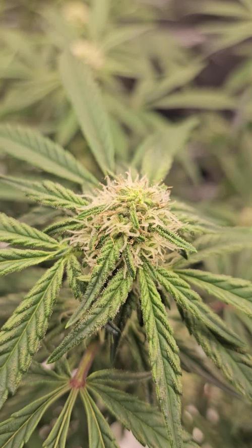
After 1st big defoliation, Plants took 4 days to recover 100%. Started eating 1 day ago. Guzzled H2O tho. 5 day, early am, plants grew all fan leaves back but smaller. Fuzz and crystals are forming. Maybe 3 weeks if I lucky. Need to stretch. Small buds rn. 4 days after post video and trichome pic taken after defoliation during early flower/mid-flower


Likes
22
Share


@dillande3
Follow
Hello, Day 47, 48, and day 49, Flower Phase , PGK, Everything looks fine, seems ok for the moment. RH is high these days, very near to harvest and i am going to finish this grow after 10-12 weeks or Day 70-84. also gave 2nd dose of NPK (0.6-3-3).Thanks.
Week 7 Flower Phase
Day 46 - 19/12/22
Day 47 - 20/12/22
Day 48 - 21/12/22
Day 49 - 22/12/22
Likes
Comments
Share


@naughtydog420
Follow
This was my first attempt at growing autos she triggered a little to early being the reason she is so squatty but I'm sure she's still going to be just as good as a plant twice her size I most definitely learned a lot about the auto flower that I didn't know before so I made adjustments on my second auto flower that I'm growing she is about 40 inches in height and filling out nicely i'm going to let the plant pictured go another week or so to make sure she's completely done I will post pictures and video of the harvest
Likes
17
Share


@Island
Follow
Week 8 -
Dead line is coming, starting week 8 of flowering.
The routine is maintained, feeding only with water.
I think Black Jack will pass 10 weeks of flowering.
Likes
11
Share


@VagosGrows
Follow
Buds Densed up pretty nice this week and got a good amount of trickles. I’m Happy How This Week Went With The Girls.


Likes
158
Share


@Ezzjaybruh
Follow
Doggo update
Couple vids sprinkled in here.
Love doing slo-mo with frisbee throws and treats catching in their mouth.
Slo Mo vids didn’t upload slo mo so I had to go thru and edit them and screen record it just to get them in slow mo on the site but they’re worth it
Dukes ear has been bugging him and so he pins it down sometimes with the other ear straight up and he looks goofy as hell.
