
Likes
Comments
Share


@FlavoursOnly
Follow
First week under big light, and in big pots. The light is at 50% giving me an average 550umol at 2ft. The ec fans are running about 50%. Total power consumption is around 600w so costing about 0.10 per hour to run.

Likes
30
Share


@TheGourmetWeed
Follow
Hi guys!
Here we are again, closing report for week 12, or 8 of flowering, as you prefer.
This was a great week!
I'm super happy in first place, to be home again, and that gives me much more time to look after the plants, the conditions, and really take great care of the grow.
All the 5 plants had great development in their bud production and amounts. The grow tent looks small for this 5 girls and now they're struggling a little with the space.
About the feedings: I started flushing already. Might be early for many, but I don't see the need for more nutrients when the plants are looking so nice, and I'm also satisfied with it, so now just water until the end.
Curiously, the 2 shortest ones are showing signs og being ready very soon as you can see in the close up shots and video. I might give it another week and will chop them both around december 13/14
The remaining 3 ones... they're still work in progress, growing enormously big for my expectations, and filling up the empty spaces with sweet, fruity and tropical aromas in the room.
Damn it smells nice when you open that grow room.
So yeah, basically now she's just finishing up, drinking water at 6.3 to 6.5PH max, 28 to 38ppm, the temps are good, 18 with lights off, 27 lights on...
The only 'issue' this week it turning the plants a bit because they're bending with the weight. Besides that, all good.
This happy grower is done for this week, let's see what week 13 will bring us.
Thanks for reading, hope you liked the pics also.

Likes
13
Share


@Mrs_Larimar
Follow
2022-10-30
The Seed was gifted to me by my Growmie @Oimrausch
thy Buddy!
She was growing from beginning very well
Turned out to a strong well structured Plant
Buds are dense and Thick, she smells super
This Run was one of my Best, I feel iam reaching good Levels in the Growingconditions
VPD, Wind, and Fertiliser helped the Genetics to show
what they got!----
Especially to mention are the Hyphotonflux HPF-4000.
All Plants responded very well to the Lightspectrum
Likes
Comments
Share


@TyRun
Follow
Clones adventure. Mom's recovering.
Spoiler: it was a very bad idea to put the clones into such large plastic pots and flood the propagator with too much water. Yes, the environment was fine, but there was no airflow through the coco, so the cuts rotted and I had to re-transplant the clones again after cleaning off the damaged parts.
Meanwhile, the mom is happy and recovering very fast.


Likes
30
Share


@GYOweed
Follow
Just been giving her water till today, megacrop part 1 only and 1/4tsp gal pk boost.
Smells like sweet cucumber mmmmmm.
Day 25 looks good imo.
Got a clone or two of it for breeding.
Likes
3
Share


@Dysons12
Follow
Nothing much to report as it’s business as usual. Feeding every other day and water and cal mag in between. I will be feeding less and less over the course of the next week or two.
Just keeping up with everything and making sure temps and humidity is at its standard
They are coming along and can’t wait to see the end product


Likes
14
Share

@BlaKX
Follow
High Growmies leider habe ich ausversehen den vorherigen Beitrag von der Ernte gelöscht..... deswegen habe ich jetzt einen neuen GMO Auto Harvest report hochgeladen ^^


Likes
30
Share


@coyote2thick
Follow
Flushing this week with pH water for a couple of days and using GH flora kleen the last two days. She smells something amazing.

Likes
3
Share


@Luv2Grow
Follow
Day 71 - Starting off week 11 with 2 gallons of water and 15ML of Cal-Mag. She’s definitely starting to beef up and getting pretty frosty and smelly now. Had to remove a few more leaves towards the bottom that we’re dying off and just tucked the rest once she was out back in the tent.
Day 72 - Not much happening right now, she’ll definitely be ready for water again tomorrow and tomorrow’s dose will be with nutrients. Just kinda fluffed her up a bit and tucked the leaves before putting her back into the tent.
Day 73 - She was dried up again today so she got 2 more gallons of water and nutes. The smell is really starting to get strong, like a sweet funky cheese.
Day 74 - Nothing new today, just took her out for new pictures and out her back in the tent and tucked some leaves. Noticing a two branches that just aren’t getting much light so thinking about chopping them off but not sure because it could be too late to do it and don’t want her to be stunted this late into the grow.
Day 76 - All looking good now, she was a bit droopy today because I didn’t get a chance to water her yesterday but within an hour of getting water today, she perked right up.

Likes
38
Share


@ILoVeThiSpLaNt
Follow
La pianta per le bassissime temperature di fine ottobre è stata tagliata qualche giorno prima ma sicuramente la rifarò indoor. Sono molto contento e difficilmente lo sono. Ho detto tutto 😊🖐️


Likes
59
Share


@JeyGanesha
Follow
Prima settimana di fioritura,tutto procede più che bene,,anche questi altri giorni si sta riprendendo ogni giorno di più. ..
Likes
16
Share


@MarcGrows
Follow
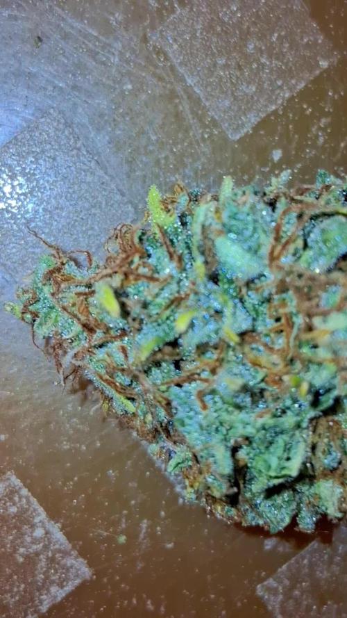
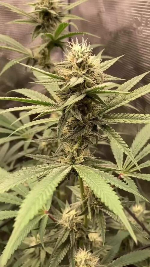
Great week. Girls finishing up and taking longer to drink. Checked trichomes and this plant is ready with mostly cloudy and a spattering of amber. Will harvest tomorrow morning and will provide weights and measures after harvest. Will dry in tent without light and offset circulation. Will keep tent naturally at 67 degrees. I will need to assist with humidifier to get humidity to around 60 RH. Perhaps a wicking cloth in a bucket? Just thinking out options.
Update: Cut down the girls and hang them up to dry. Great thick hard buds. Can't wait to see what they smoke like, but it is going to take time to dry. Thanks to everyone that helped to make this a successful harvest. I will weigh the harvest before starting cure after a closer trim.

Likes
Comments
Share


Likes
13
Share


@NordicClosetGrower
Follow
Finally received a couple of humidifiers to test from the local shop. These shitty low cost humidifiers will not really do, I can only have them on non stop until the water runs out. If I connect them to a sensor that switches them off, it won't go back on because it doesn't have a mechanical switch, I have to press on each time.
Still, let's see if this helps my poor dry leafs a little bit
Likes
35
Share


@madlangs
Follow
Looks like I have been under feeding and thinking the plants was going pale because the light was to low. Now feeding full strength and lowered the light a bit today
11.10.21
Dg 4L
Wc4L
Gb4L
Chem4L
Cream.4L
Gave all full strength week 2 flower with fish mix. Recharge mammoth p. Silica 1ml/L calmag 0.5mlL. Ec 1700 ph ph6.4
Hubba.4L
Orange. 1 3L
0range 2 4L
Gave all week 1 flower with fish mix. Recharge mammoth p. Silica 1ml/L calmag 0.5mlL ec1500. Ph6.4
13.10.21
Dg. 3L
Wc. 2.5
Gb
3L
Chem. 3
Cream. 3
Hubba 3
Gave all week 2 flower apart from hubba got week one flower full strength silica 1ml/L. Calmag 0.5L/L ph6.4 ec1700 hubba ec1500
15.10.21
Dg. 5.5L
Wc. 3L
Gb 4.5
Chem. 4.5
Cream 4.5
Hubba. 3.5
Orange. 1 4.5
0range 2 5
Gave all full strength full strength week 2 flower gave grow instead of fish mix
Calmag 0.8ml/L for some reason ec was ec2.3 so added more water to bring down to ec1.9. Watered dg with it and added more water to bring down to ec1.8 ph6.4 for rest of the plants
Run off
Wc ph5.7 ec3.4
Hubba. Ph5.9 ec3.0
Orange no2. Ph5.9 ec3.6
Orange no1 ph6.0. Ec3.8
Chem. Ph5.8 ec3.1
Dg ph5.7 ec4.5
Cream ph5.9. Ec4.3
Gb ph5.5. Ec4.3
15.10.21
Dg 60cm
Wc 54cm
Gb 58.5cm
Chem
66cm
Cream. 61cm
Hubba. 50cm
Orange. 1 28cm
0range 2 38cm
16.10.21
Spectrum king 90cm
Lumatek 70cm


Likes
24
Share


@BillMonroe
Follow
Lord have mercy on my plant 🙏
After finding some spider mites last week I have released predatory mites. Phytoseiulus persimilis. They came in a bag with leaves and you put those leaves on the canopy.
Spray the room with spider mite insecticide.
I found no more spider mites and no increase in damage. Until now the damage has been very minimal, compared to pictures I see online. Just a few white spots on a small number of affected leaves. Never saw any webbing, neither on leaves nor buds. Regardless I am treating it like a big infection
Oh yeah, it's almost ready, I guess? All I can think about are little spiders. Thanks to everyone for watching, and your tips and advice.
