The Grow Awards 2026 🏆 


Likes
Comments
Share
@Secretflower
Follow
Hello my friends,
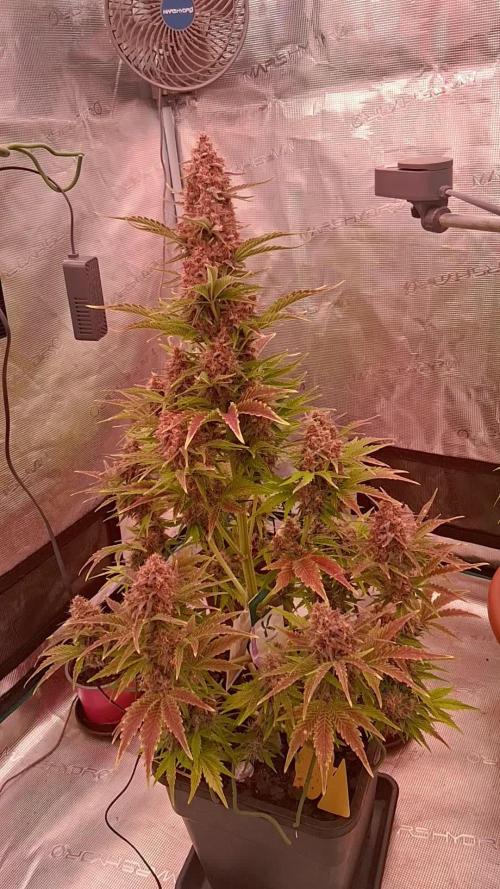
...May 30 , 2022....Day N°79..
...Flowering day N°23...
My three Feminized Watermelon Candy are fine, there are beautiful, flowers smells awesome.
I feed them with the Hybrid powder and some Boster from Green House Feeding Nutrients. I gave them some CalGreen from Metrop, the best Cal-Mag of the market.
They are under a MarsHydro TSW 3000 at 60% of power and at 30cm of the canopy.
www.zamnesia.com
www.mars-hydro.com
Thank you very much for passing by.
Wish you only the best with your green projects, peace.
See you soon 💨💨💨


Likes
5
Share
@jojopfoh
Follow
She is just bulking up now. I am cutting back the bloom as the tips are burning.

Likes
16
Share
@DeepSouthDank
Follow
Today marks the beginning of my Hella Jelly pheno test run indoors. I've selected 7 out of 9 of the plants to re-test indoors from the outdoor mini pheno hunt. Unfortunately, I didn't post that grow! Life got hectic.
Clones were taken 7 weeks ago and have been vegged for five weeks, the plants will be going into flower cycle tomorrow so we can figure out which ones are the true keepers. Some of the plants are a little behind, but we have taken more clones and we want to see what we are working with now before some are to big.
A few of the Pheno's showed amazing potential outdoors and I am eager to see their true capabilities under perfect environmental controls.
Plants are currently fed once a day by hand. The multi drip feed system will be installed in the coming weeks.
Plants are fed at 800 EC and we will slowly work our way up to 2000 over the next few weeks.
Installing some UVB bulbs in the next week or two.
I am using some techniques to keep the stretch minimal to encourage strong plants with dense stacked buds.
I'm excited to see how these plants develop over the coming weeks and
I'll update my diary weekly with progress!
Happy Growing
I have added some pictures of the pheno's from the outdoor grow

Likes
69
Share
@Hempy_The_Kid
Follow
5/12/21 Week 1 of flower 10 of life. Checked fluids 6.01ph, and 390tds. Plant seems ok were I split stem. Will give dose of recharge later this evening. Now on a 12/12 cycle.
The watering volume per hour is not letting me go over 2.6 gallons per day. My pump is recirculating water at 200 gallons/hr it runs 15 minutes 2x day. So 50 gallons 2x day or 100 gallons per day.
5/13/21 will start foliar feeding tonight at lights out. Using TPS Canopy boost. Will use for 3 weeks every 3-4 days. Changed water to bloom nutrients. Ph 6.01 tds 710.
5/15/21 ph 6.18 tds 570. Will get dose of recharge this evening. Plants leaves are almost touching side to side in tent.
5/16/21 added water (4 gallons) can't believe she went through that much in 3 days. Ph 5.85 tds 689. Will get a foliar feeding tonight at lights out.
5/18/21 changed nutrients water. PH 5.96 TDS 680. Leaves of plant are now touching all 4 walls of 4x4 tent.

Likes
29
Share


Likes
57
Share
@Secretflower
Follow
Hello my friend,
...June 14, 2022..Day N°84...
...Flowering day N°38...
My Feminized Tatanka Pure CBD is beautiful and fine. The flowers developing very good.
I gave her water with a tablet of Easy Bloom Booster from RQS Organics Nutrients.
She's under a Mars-Hydro Tsw 3000 at 70% of power and at 25cm of the canopy.
www.royalqueenseeds.com
www.mars-hydro.com
That's all for now my friends, thx for passing through here.
Wish you the best with your green projects.
See you soon..💨💨💨
Likes
30
Share
@McGrowin
Follow
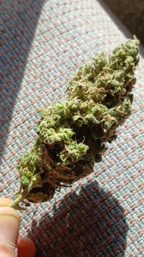
72 grams of dried and semi-cured creamy buds. smooth smoke already despite the need to drop humidity below 59% fully. not much larf surprisingly but will be fun to see how much hash each strain gets considering others in the tent had way more.
Harvest: somewhat early, but solid nugs and yield - easy trimming and quick to dry compared to other leafy varieties
terps: creamy dough mostly with hint of sweet cinnamon sugar vanilla - just like the pack says, but still not impressed really
Drama about these seeds: got store credit on the pack of 3 seeds just because of website change to outdoor only after purchase date. excited to grow other HSC strains next year.
Likes
9
Share
@I_Identify_As_A_Dan
Follow
Week 6 for Black Lebanon by SSSC,
Like i mentioned on the previous Sunday update, she was transplanted a day after the post. I was planning on not giving water for the first week to let the roots do some growing.... the rain gods had a different plan 😂 Its been storming rain ever since day after transplant now 5 days i think 😂😂 So that didn't go to plan but hoping no rain next week fingers crossed.
I also think i topped her again some point last week. Cant wait for her roots to get moving in the new home.

Likes
18
Share
@BlaKX
Follow
High Growmies, die BPP hat in der Woche nochmal ordentlich an Masse und Harz zugenommen! Auch die Terpene kommen jetzt voll zur Geltung.
Habe mit meinem neuen Smartphone Mikroskop 300x die Trichomen gecheckt und festgestellt dass fast alle Milichig geworden sind auch vereinzelt Bernsteine aber noch hier und da durchsichtige.
Da ihr Durst noch nicht abgenommen hat werde ich ihr noch eine Woche mehr geben um die Trichomen Reifen zu lassen und Festigkeit zu steigern.


Likes
44
Share
@DutchFarmer
Follow
I found an awesome strain with four seeds and tested three of them. The smell was pure vanilla gas—super stinky and strong. Phenotype 1 was the best, with amazing density and a frosty look. It was a heavy yielder too, giving over 7 ounces from a 1-liter COCO. The terpene profile is incredible, tasting just like cookie dough. Would I grow this strain again? Definitely! I even saved a clone. I'd rate it a perfect 10/10

Likes
93
Share
@Natrona
Follow
FBA2502 Plants 1-6
Week 7 May 11– 17
FLOWER 3
Nutrients :
Micro 25 ml
Gro 10 ml
Bloom 30ml
CalMag 30 ml
Recharge 10ml
Green sensation 16.5ml
Power buds 7.5 ml
5/11 PH 6.56, PPM 1560, temp 69
2ltr each
5/12 PH 6.31, PPM 1120, temp 68
1 ltr each
5/14 PH 6.66, PPM 1280, temp 70 1 ltr each
5/15 PH 6.66, PPM 850, temp 72 1 ltr each
5/11 Defoliation, feed, pics & videos
I made 4 gallons, used 3 to water then poured the runoff back into the rest in the bucket. The next feeding, I added a gallon of tap water to residual and ph to 6,31 temp 69 & PPm 1120. Did the same on 5/14 and 15 using up the remainder of the feed solution.
I must monitor and control the tent environment manually. For the past month, the humidifier has been an issue putting out way too much humidity; often leaving water in the bottom of the tent. In addition, the temperatures are also out of control. I added an AC last week to keep the temperature below 80. I finally determined the exhaust vent is also broken and has been for some time.
I have been giving good reviews on the AC infinity system. However, during the time I’ve owned this tent (2 yrs), I had to replace the lights twice, fans, humidifier and now the vent twice also. Everything but the tent containment has been replaced. AC infinity does stand by their products, but they are in California, and it takes at least 2 weeks to receive the replacement. This is extremely frustrating at the critical transition to flower for my equipment to fail again. For the time being until a replacement arrives, I pulled the 4” exhaust vent from my 3x3 and hooked it up. I have the AC pushing air in and the exhaust pulling air out. This should create a neutral pressure tent environment-neither positive nor negative. I do have 3 circulating fans in the tent to move air around the plants.
It looks like growth spurts have slowed and plant height has been reached. Now is the time for buds to fatten and frost up. All of them (6) are short remaining less than 3 ft. They range from 15 to 31 inches. The tallest #4 &5 and #6 have very light leaves and appear nutrient deficient compared to the shorter phenotypes. Pictures taken later in the week show fading on a few the older leaves. All are receiving the same solution and are in the same soil blend. Moving the gals in and out of the tent for their glamor shots, I bent one limb and snapped another. The bent one I taped with a splint and the snapped one I medicated manuka honey and wired it back to the main stalk.
Leaves look like sativa leaning. I hope FBA2502-2 is a good daytime. I’m getting excited to see what develops in the next month or so. News Break: I’ve been informed that 2502 is FastBuds New Frostbanger and is indica for nighttime. I’m disappointed that I didn’t get a sativa to test because I really need a daytime strain. The 2 Blue Cheese plants I just grew are also nighttime.
Upon opening the tent, a faint odor was detected, and some stickiness was observed during the process of defoliation. Pictures of the puffy buds even show differences. #4 &5 buds hairs are different from the others being crinklier and the others are straighter. Some like #2 &5 are showing frost on the leaves. Wow at the beginning of flower to have that much frost. They are living up to the new strain’s name.
I’ve noticed that when I water, sometimes water shoots out of the side holes I the air pots. I will pack the soil firmly into the sides for future runs.
Measurements:
#1 15”
#2 20”
#3 17”
#4 30”
#5 31”
#6 24”
Your likes and comments are appreciated. Thanks for stopping by.
Growers love 💚🌿
💫Natrona💫


Likes
21
Share
@DeeDeeKushner
Follow
These videos are from yesterday and we’re on day 5 of flower. Been MIA trying get this girls back to being healthy. I think they’re pretty healthy these days for sure. I did a bud leaf strip yesterday and some super cropping as well. This strain takes 8 weeks to flower completely. Looking forward to seeing how they turn out! ✌️🏾


Likes
4
Share
@kdifiori_
Follow
It's harvest time for Pineapple too. This little one has grown very quickly since the beginning, living up to her name and surpassing all the others born with her in height. She caused no problems, completely indifferent to everything going on around her. Beautiful to look at and wonderful to smell, completely covered in trichomes. It's like smelling a pineapple! So fresh, so tropical!
Processing
Likes
10
Share
@LockDownGrow
Follow
This lady started too pile on frost from the first week of flower, something tells me this lady is going too be fyah

Likes
2
Share

Processing
Likes
10
Share
@PisBaked
Follow
Week 5.5 for PE, GSC (small one), & both GG's. Week 6 for everything else!
Everything looks great other than the one GSC.. What I thought was light burn is apparently nute burn.... Run off PPM is about 100ppm higher than the rest... Gave the whole crop a little flush, and gave them half strength feedings for 2 days. Obviously the burnt tips won't recover, but it doesn't seem to be getting worse. Right now she's at a whopping FOURTY EIGHT inches... She is the most developed, and has officially begun producing buds. Scent def. resembles GSC, no question at all.
Next best is the LSD25, growth is amazingly healthy, and is starting to form buds.
Everything else is doing great, but going at pretty normal speed compared to the LSD & GSC
Healthy white roots are exploding out the bottom of every pot; something I've never seen growing in soil.
