


Likes
Comments
Share

@GrowBro1981
Follow
Hallo liebe Growfreunde 💞
Diese Woche kamen die ersten Problemchen ?!
Mein pH Messgerät hat falsch angezeigt und ich habe es erst nach dem 2. Gießen bemerkt. (Deshalb ganz wichtig: "IMMER WIEDER KONTROLLIEREN UND KALIBRIEREN"!!!) Blatt Nekrosen wurden bei einigen sichtbar, da der pH bei 5.5 war, konnten die Pflanzen die Nährstoffe aus der Erde nicht verarbeiten, deshalb wirkt es wie eine Überdüngung, aber ich dünge nicht, deshalb weiß ich dass es ein kleiner pH Lockout ist.
Glücklicherweise haben sich 4 von 6 sehr gut davon erholt💪🤗. Bei einer Gorilla Zkittlez und einer Special Kush wird sich kommende Woche zeigen ob sie bleiben dürfen oder nicht. Evtl. Steht auch das Topping an ,wenn die Pflanzen wieder fit sind.
In diesem Sinne wünsche ich euch einen schönen Sonntag und eine erfolgreiche Grow Woche ??💪. Bis nächste woche meine lieben 💞💞💞


Likes
7
Share


@chronicster
Follow
10/29: Watered 2 gallons in each plant. Pulled branches down and out to spread as wide as possible. Things are looking good, pulled a few dead leaves and it has a couple that have brown on the tips, but past experience tells me that it will pass and they will be fine. I'll keep an eye and determine if anything needs to be done.
11/2 - They lost the friend that was finishing up and gained a new friend who is in week 1 flower. This one is another non-diary plant that I began before being on here.




Likes
Comments
Share

@HOME_GROWBOOK
Follow
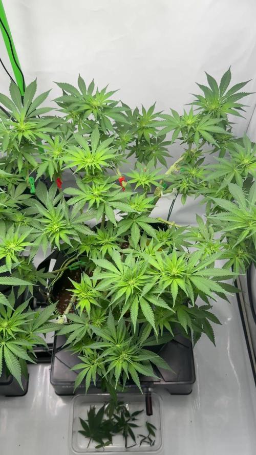
Der Stretch neigt sich dem Ende zu und das Netz wurde entfernt – beide Pflanzen stehen jetzt stabil und frei im Raum.
Nach dem Lösen des ScroG zeigt sich deutlich, wie kräftig die Struktur aufgebaut wurde:
dicke Stämme, starke Seitentriebe und ein gleichmäßiges, volles Blätterdach.
24K Gold reagiert mit einem beeindruckenden Stretch-Finish. Ihre Haupttriebe sind kräftig und leicht über dem Restniveau, perfekt verteilt für maximale Lichtausbeute. Die ersten Blütenansätze sind klar zu erkennen – kleine weiße Härchen schießen an allen Spitzen hervor.
Gelato 33 bleibt etwas gedrungener, aber sehr kompakt und buschig. Ihre Top-Sites sind gleichmäßig verteilt und zeigen ebenfalls die ersten Pistillen. Das entfernte Netz hat ihr Raum gegeben, sich frei nach oben zu entfalten.
Eine gezielte Entlaubung wurde vorgenommen, um die unteren Bereiche zu öffnen und die Luftzirkulation zu optimieren. Das Laub ist tiefgrün und gesund – keine Mängel oder Verbrennungen sichtbar.
Düngung & Werte:
• 💧 Canna Coco A + B → 6 ml/l
• 📈 EC: 1.6 pH: 5.8
• 🌡️ Temperatur: 25–26 °C
• 💧 Luftfeuchtigkeit: 50–55 %
• 💨 Umluft: stark & konstant
• 💦 Tropfbewässerung mit kontrolliertem Drain
Der Fokus liegt nun voll auf Blütenbildung – die Pflanzen stehen stabil, haben den Stretch abgeschlossen und beginnen mit sichtbarem Blütenaufbau.
Ein sehr kräftiger Start in die Hauptblütephase!

Likes
35
Share



Likes
22
Share


@Damonkey
Follow
Last 2-3 weeks of the grow period now, expecting the COP to finish up first and I’m guessing the PSS is going to push it to 10 weeks, at least I hope so, would be awesome to have a fairly nice yield from that one. It looks and smells so good just need a bit more swelling inna da buds. All looking great. However got a slight nute burn to all plants from one of the last feeds. Only very slight and it was picked up quickly. The following feed consisted of Tripple F and molasses soup, holding back all other nutes until the following feed. Problem seems to be sorted.
The grow becomes a little more of a challenge day by day, the weather here is worsening meaning colder and more humid day by day, my cramped circumstances don’t help the situation either. The hum coming from my room now is also a little to loud and when everything’s running at quiet times can be Heard, when it really shouldn’t be, although I don’t have problems with that at the moment who knows who might hear it at the wrong time🙄 oh spring hurry along please.


Likes
16
Share


@Foggedaboudit
Follow
10 weeks total for the first phenos and a bit more than 11 for the last one! I could have easily gotten double the projected yield on these had I not neglected them but whatever I pulled is rock hard, dusted in sugar and smells absolutely stupendous! You did it again FastBuds!

Likes
34
Share


@Brick_Bazuha
Follow
Неделя прошла замечательно, от дефецита избавились точно, шишки набухают каждые 2-3 дня заметно для глаза) запах божественен, правда отдаёт свежей кожурой апельсина) осталось почти немножко)
В конце видео есть с правого боку два клона с этой вдавы, но про них это другая история)

Likes
34
Share


@gottagrowsometime
Follow
Welcome: thanks for dropping by. Drop a like so I can check out your grows, or not, I'll leave it down to you.
So, the fun begins with Kosher Cake. Have 6 in the ground FB seeds in the ground only 1 kosher. The 2 Strawberry Gorillas are still not there with germination.
Have started these on the 3 nutes above. These are best known as Voodoo Juice, tarantula and Piranha. These versions are from the ppl who hold the pattens and make a lot of ANs formulas. They have just improved upon most of the AN line up but they kept these the same. As they are tried and tested. Going to be running this lady on the full 19 Terra Power Line up. You can check these out on led-grower.eu/terra-power/ and read up on them for yourself. I'll be adding what's in them as for these 3 if you are interested look up Voodoo, T&P. Hopefully soon you'll be able to click on them and look em up when they join GD in the next wk or so.
Happy with how the nutrients are holding a stable PH at 6 and these aren't even their PH perfect nutrients. So no PH - + needed.
Sprayed each plant expect 1 gorilla zkittlez, for that 1 I used ANs line. These recommend 1ml/l for seedlings, AN I used 1.5ml/L. I use to use 2ml/L but they are seedlings so it's enough. Just a waiting game now. Will update as soon as these ladies start a popping.
I laid a damp towel around the pots hopefully it'll up the RH. (Don't soak the towel it needs to be able to dry easily and in turn increase RH)
Sprayed each 5 plants with this line up to start them off. As soon as leafs show I'll start them on a very very light feed. Will feed again in 2 days with the same mix. Another 100ml 1ml/l of each 3ml/l total 627ppm 6ph 21c temp. Have the smart intake fan at no 4 on 100% of the time. If I can't get desired RH after a day or 2 I'll turn the intake fan on 20/20 on/off and it'll make the RH be above 70% by doing this alone in a small space like 70x70. I really don't think I'm gonna have an Issue with low RH, but I have a feeling I'm gonna pay to have a low RH environment.
30/4/22 VD1. Have started her on jungle juice trio and a mix of terra Power enzymes and all there bud/resin enhancers will be used in conjunction with half strength recommend jungle juice dose. The feed is so low I've made up a 1l mix and that will do this and the 2 banana purple punches also. And I'll move that up to 1ml/L x3 of the jungle juice products next week. But for this week what's up above will be given every 2 days or when needed.
1/5/22 Slow growth on VD2. She's the runt most are reaching 2.5cm 3+ nearly on 2. Let's see what happens she's out of the ground and showing fan leafs, yday she had a crusty head. So all in all, going well. 6-8 FB seeds are up and thriving.
2/5/22 She's picked up fine. Got some height to her. Will continue the same course and watch her grow. Will update once more on on size and daily pics.
4_5_22 All Jungle juice&Terra Power grows are showing deficiency signs. I'm going to push up the EC this week I think. I do not want to stall them but I really think EC will be high in jungle juice because of the micro it has SO MANY TRACE nutrients it's crazy and it sends the EC sky high but there's so much of little bits of this and that I really think it can handle a higher EC. I'm going to 2x it or up it by a 1/3 for their watering 2moro as they were only fed yday. I've added biobizz calmag to help as my tap water is lacking cal mag when it comes into summer or so it stays on our water reports. It drops by 20%.
6/5/22 So, it's begun. I've created a negative pressure environment, 1st time, let's see if it makes an impact. On wk 3 onwards I'll be running the intake extraction for 5mins every 30mins and I'll have 2 co2 bags up. If the RH becomes a problem or temp I'll put the extraction/intake back on 100% of the time. The extraction fan/intake is set at a ratio of 2 to 1. (100% extraction 50% intake) making a negative pressure environment, best way to show you have done this, the walls in your tent should bow in lightly.
Same Day. Okay, so this my last update. Going to run fans the same right up until flower and then I'll be sealing up my tent and adding 2 co2 bags. And will be adding a dehuimifier and setting at 45% And it checks every 30mins and if it's gone up it'll turn back on and bring down to 45 or set RH, so no co2 will escape. The temp will be bordering 30-33c and then when they go into their rest window they'll be transferred to to a negative pressure environment with low temps and 45-50%RH. I'll only be able to do this for the 3 weeks of pre flower. And maybe some more, it depends how hard the dehuimifier will be working, but i want dense buds and only way to improve that is by introducing Co2 and then a negative pressure cold environment for terps. Then I'll be putting them back into full negative pressure environment and the Co2 will be useless, again all depends how it's gonna affect my bottom line. That and I only have 2 intake fans. I'll be going into detail about my Co2 adventures the week before I'll be prepping them to be able to withstand high temps, so I'll gradually bring up the temps the last wk of veg 28c max and that way when I push it to 30-32c in pre flower she won't stress as much
Thanks for reading, this will be it as for updates for this week other than her final 7th wk day. Will adjust EC 5th higher or so.
gottagrowsometime

Likes
3
Share

@RicoGrow
Follow
Hello,
I hope you're all doing well.
I'll consider this week a complete failure.
When I repotted my plants, I added mulch, but it was only 3 cm thick.
As a result, my plants were miserable for four days because it was too thick.
The soil was cold and remained very damp due to watering during repotting.
It smothered the fallen leaves, some of which even turned yellow. It seemed like a magnesium and calcium deficiency and an unbalanced pH. I re-evaluated my approach and, most importantly, added a light layer of mulch.
They've recovered well. It just goes to show that a small mistake can teach you something new every day.
This week, I added:
2 g of Epsom salt:
This powerful mineral is pure magnesium sulfate, one of the main components of chlorophyll produced by plants.
It's a chelating agent: it allows the solubility of other substances (such as molasses, humic and fulvic acids, neem oil, and spirulina) and acts as a heavy metal sequestrant.
5 g of Micronized Wild Horsetail Spray: silica, defense
5 g Fish Hydrolysate: amino acids, proteins
ppm 600
pH 6.5
Wishing you all a successful cannabis grow!
****************************************************
Bonjour,
J'espère que vous allez tous bien.
Cette semaine, je la compterai comme nul.
En fait lors de mon rempotage, j'avais fait un paillis, mais il était de 3 cm.
Du coup mes pieds, on fait la gueule pendants 4 jours, car il était trop épais.
La terre était froide, rester très humide dut à un arrosage lors du rempotage.
Elle étouffait les feuilles tombées, elles ont même jauni pour certaines. Comme un manque de magnésium calcium et un PH non ajusté.
Remise en cause et surtout un paillage léger.
Elles sont bien reparties. Mais comme quoi une petite erreur et on apprend tous les jours.
Dans ma terre cette semaine, j'ai rajouté.
2 g de Sel d Epsom:
Ce puissant minéral est du pur sulfate de magnésium, l'un des principaux composants de la chlorophylle produite par le règne végétal.
C'est un chélateur : il permet la solubilité d'autres matières (comme la mélasse, les acides humiques et fulvique, l'huile de neem, la spiruline) et agit comme un séquestrant de métaux lourds.
5 g de PRÈLE SAUVAGE Micronisée Aspersion : silice, défense
5 g HYDROLYSAT DE POISSON : acides aminés, protéines
ppm 600
PH 6,5
Je vous souhaite à tous une bonne cannabiculture
👍👍👊😁😁😁


Likes
8
Share

@Smoking_Joe_Frazier
Follow
02.08.2026
Hi Growmies,
welcome back to week 8 of my Fast Buds Auto Grow!
Weather and Conditions
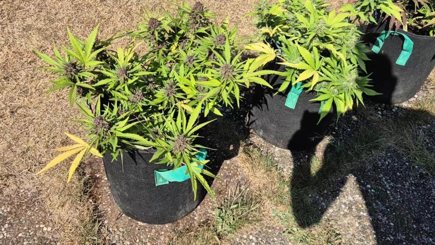
The weather has been almost perfect for the girls, with plenty of sunshine, high temperatures and still no rain at all. If these conditions continue for another two weeks, I might make it through the grow without any major issues. Fingers crossed!
Unfortunately, my rain barrels are completely empty by now, so I've had to switch to tap water.
Growth
The girls have completely stopped stretching and are now focusing all their energy on swelling their buds and producing more resin.
Papaya Cookies
No more side branches are developing, but the buds are getting noticeably longer and fatter every day.
Her smell has become incredibly intense. With the right wind, you can already smell her from the front of my house. Definitely not the best strain if you have neighbours with sensitive noses.
The colours are absolutely gorgeous as well—a real eye-catcher. I estimate she'll be ready for harvest in about two weeks.
Tangie
When it comes to bulking up, she has now overtaken Papaya Cookies with ease.
Just like in my 2024 grow, she's producing long, candle-shaped colas. The genetics seem to be incredibly consistent.
The aroma reminds me of freshly peeled oranges, and I absolutely love it. I think she'll need around three more weeks before she's ready.
Apricot
Apricot is also showing some beautiful colours, although she'll probably end up being the smallest yielder of the three.
But... she easily has the best aroma of them all. In my opinion, she even smells better than Papaya Cookies, although not nearly as overpowering.
Fast Buds really created a beautiful terpene profile with this one. Her buds aren't as large as the others, but they already look as if they've been coated in thick sugar. She's by far the frostiest girl in the greenhouse.
I have a feeling she's going to deliver a seriously potent smoke.
I also expect her to finish in about two weeks.
Feeding
Nothing has changed this week. I'm still feeding T.A. TriPart as before.
Over the next week I'll switch Apricot and Papaya Cookies to T.A. FinalPart, while Tangie will stay on TriPart for another week because of her longer flowering time.
Pests
Still nothing to report.
Looks like the grow gods are on my side this year. 🤞
Final Thoughts
Overall, it's been a solid—though not spectacular—grow so far.
Missing a feeding while I was on holiday probably cost me a bit of yield, but that's perfectly fine. The vacation was definitely worth it.
Now it's all about finishing strong and letting the girls ripen to their full potential.
That's it for this week, my friends.
Catch ya next week!
Smoking_Joe_Frazier
Likes
35
Share


@ThatoneAKguy
Follow
Cruising right along. I bent over the stalk and had to take a couple of the massive leaves off and it’s back up to 12”. I love my new system and the easy maintenance makes it even better.
Likes
15
Share

@masterofsmeagol
Follow
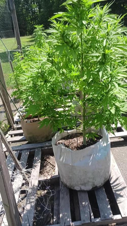
8/3 Went over to treat the 10th planet that needed water with plant doctor around 10:30. I was in the garden at 6 but didn't have much time. That plant seems to be curled up or "droopy". It DID need water. I defoliated a few sep leaves off it but I didn't see any today. I guess I don't ever really see the leaves "up" or praying like I do with the rest of tge plants. It's starting to flower like the rest though. Today is the day everything is supposed to get its second or third plant doctor dose but I decided to wait until they need the water. I don't think 24hrs will mean much. I've been staying on top of defoliation but I'm seeing some pillar damage. I'd like to spray bt but what my buddy said about spraying with septoria is still in my head. Maybe I can use citric acid. That way it won't "wash away" spores it will just eat them. At least I think. And I hope lol. Birds get most of the pillars but I hand pick some too. I need to pick up nutes. Wondering if I should just get grow big again. I don't use much but plants are switching to flower and leaves are fading. I have big bloom and kool bloom bit neither have nitrogen. I still need some sort of nitrogen. Especially since that could be a deficiency. Not sure if it's the transition or a condition with the sep. I see some interior small leaves bleeding out and some dying. I'm wondering if what I thought was earwig damage years prior was really a combo of sep and they just ate the evidence. I'm super grateful though. I've got some resilient girls and they're doing well. The special kush in flower is gorgeous as well. Looks like I'll have an early plant. Short flowering time on this strain. Smells pungent.
8/4 Treated everything in the garden with plant doctor except the 10th planet I got yesterday. I need to take a closer look at that plant. It rarely looks "happy" like my others. I'll check it again later. 3tsp per gallon and 1gal a plant. The 10s got a half gallon. This seems to be working great. I have some defoliation to do on the tenth planet I was talking about earlier. Everything is in some form of flower. The flowers on that early special kush are getting bigger. I'll update this later.
UPDATE: WENT BACK OVER AT NOON TO TAKE A LOOK AT THAT OTHER 10TH PLANET TO SEE IF IT PICKED UP AT ALL. IT DID. IM GLAD I CAME OVER TO CHECK THOUGH. MAKES ME FEEL BETTER. THE OTHER 10TH PLANET LOOKS HORRIBLE O. FILM FROM ONE ANGLE LOL BUT ITS REALLY A GREAT LOOKING PLANT AND IM USUALLY FILMING AT LIKE 6AM. JIST WANTED TO STICK UP FORCMY GIRL AND HER BAD HAIR DAY. ITS BEEN HIT AND A FEW OF HER LEAVES ARE TACOED. SHE GOT HER FIRST "PREVENTATIVE" DOSE OF PLANT DOCTOR WHICH WAS HALF A TSP/GALLON. IT REALLY SEEMS TO BE DOING SUBSTANTIALLY BETTER. EVERYTHING IS. IM HAVING TO LOOK HARDER AND AM HAVING TROUBLE FINDING LEAVES EFFECTED BY SEPTORIA. KNOCK ON WOOD. IM EXCITED. THIS IS GOING BETTER THAN I THOUGHT. IM STILL GOING TO BE MONITERING LIKE I USUALLY DO.
8/5 Watered the 10th planet I dodnt get yesterday. Looked and didn't see any sep leaves. Phone was dead so I vouldnt do a video. Went back over at like 130pm and did a quick video and checked out the girls. Everything is looking good and transitioning well. I WANT to use a diy Dr. Zymes with citric acid as I've noticed a few pillars. Only reason I've hesitated is because my buddy said not to spray it. But if what I spray it with "eats" it I would think it's ok. But what do I know. Birds get a great deal of them. I watch them on the camera sit on the cage and wait for one to stick it's head up before snatching it up. Still need to get either more grow big or a bloom product with nitrogen. I was able to defoliate that other 10th planet of anything suspect. It's hard work but it's worth it in the end.


Likes
6
Share


@GermanBoy69
Follow
Die Pflanze hat einen Wachstumsschub in die Höhe bekommen. Der PH-Wert ist jetzt konstant bei 5,5. Habe nochmal Dünger zugegeben. Der EC-Wert ist mit 2,3 glaube ich etwas zu hoch, aber dagegen mache ich nichts. Die anderen Pflanzen im Turm gedeihen prächtig.
---------------------------
The plant has had a growth spurt in height. The pH value is now constant at 5.5 and I have added more fertilizer. I think the EC value is a little too high at 2.3, but I'm not doing anything about that. The other plants in the tower are thriving.

Likes
5
Share


@TheGreenDisciple
Follow
Day 80 ~ Plants are looking and smelling great. I have been keeping an eye on the trichomes for the first plant and she looks like she will be ready right after the harvest moon! So I am excited to harvest her on Friday. While plant 2 is still another week and a half to 2 weeks away!

Likes
12
Share


@Skunkiest_bender39
Follow
The fimming does what I wished. The single headbud, „twin‘ed“ into 3 headbuds. The fimming also stops the vegetation, but most times luckily only the node of the headbud! Then does the twinheadbuds build a roof-crown with the other nodes.