
Processing
Likes
Comments
Share


@Wicked_Stix
Follow
This auto ghost of is at day 21 and she has been growing like crazy since she broke the surface. She has the potential to be a real work of art if I can keep her happy. I upped the maxigro to .5 tsp per gallon and started them on a little armor si so they can build some strong branches. Still running them on 24 hour light and 24 hour irrigation. They are going to stay under the blurple lights for a few more weeks until I can get them in the big room under the quantum boards. I'm hoping they will flower around 4 weeks but my autos seem to enjoy staying in veg so we will see what happens.

Processing
Likes
Comments
Share


Likes
Comments
Share

@420DeepGrow
Follow
📆 Semana 12
Las A5 Chaze encaran sus últimos días antes del corte.
Con el lavado de raíces completado, la planta ya no empuja crecimiento, sino que concentra todo en madurez, aroma y resina. La estructura se mantiene: la de puntas secundarias termina de compactar, mientras la de cola principal conserva su porte clásico, firme y bien definido.
Los tricomas mayoritariamente lechosos, con toques ámbar en las zonas más expuestas, confirman que el punto óptimo está muy cerca. El perfil terroso–especiado se presenta ahora más limpio, profundo y persistente.
🌿 Última semana: estabilidad, riego mínimo y paciencia.
La planta se despide afinando cada matiz antes del corte.
¡Seguimos creciendo fuerte 💪!
Likes
2
Share


@TreeFruitsDe
Follow
I took this video in the 6th week of flowering.
I only used the fertilizer once in the second week of flowering 3g/L.

Processing
Likes
11
Share


@LaughingDragon
Follow
This bugger neatly drank 3L of the solution I had prepared for three weeks!! there some serious growth happening. Defoliation was a good idea at this point to reduce the amount of wasted nutrients and increase the flowering priority.
Had to share the last "video" to show my appreciation in learning about LST here on growdiaries, those are pepper plants.

Likes
26
Share

@HinduGod
Follow
WOW LOOK AT DAT!!! DIS PLANT HAS HAD IT ALL!!! YOU CAN SEE DA FROSTBITE DAMAGE!!! DER IS NOTHING WRONG WIT DAT!!! DIS IS A LATE WINTER EARLY SPRING GROW!!!! IT PROVES DAT YOU CAN GROW AN AUTOFLOWER IN SUBOPTIMAL CONDITIONS OUTDOORS!!!! DIS IS NOT ABOUT YIELD IT IS ABOUT GETTING SOME GOOD SMOKE IN DA WINTER SEASON OUTDOORS DIS IS VERY VERY GOOD!!!!
PLEASE COME AGAIN!!!!
Likes
26
Share


@pifflestikkz
Follow
Day 17
17/07/24 Wednesday
De-chlorinated tap water pH 6 with calmag 5ml -5L.
Very humid week, noticed a green turning colour on top of soils where the humidity has been far to high.
I have installed door netting and recently left open all day and night now so they can condition over night in colder temps.
All are doing well, one overdose slightly behind and looking ABIT swifted. But she'll come around 💪💚
Day 19
19/07/24 Friday
Lite Feed today, 250ml each pot small run off.
Seeing excellent start to these babies. Let's get it 👌💚
Day 21
21/07/24 Sunday
De-chlorinated tap water pH 6 with calmag 5ml to 5L.
Watering in 1L each day from now.
Updated video

Likes
31
Share


@Chubbs
Follow
SuperSativaSeedClub
PurplePunchOG Auto
Week 6
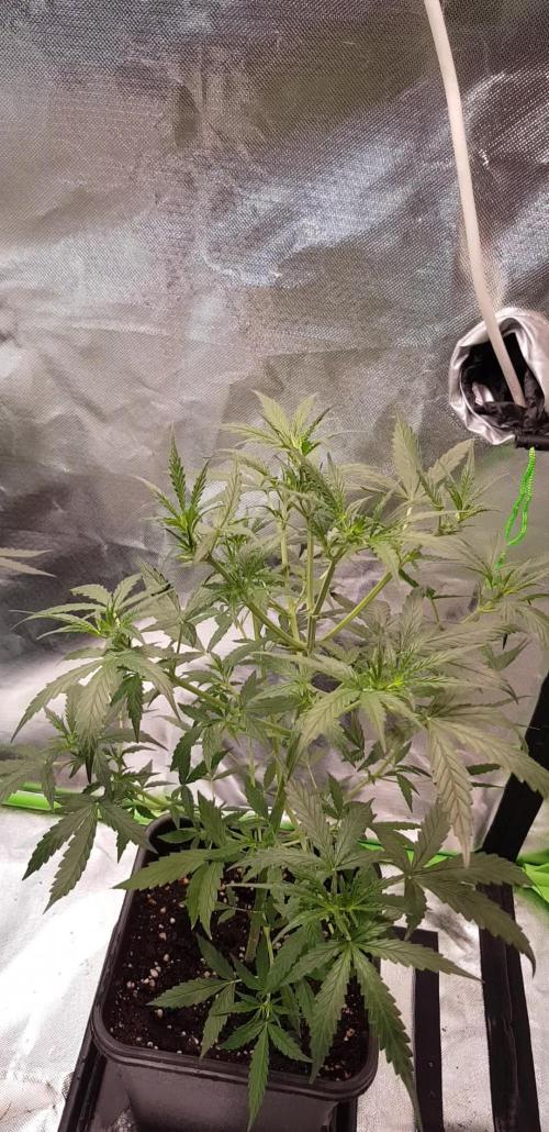
Weekly update for these three little ladies. They still haven't gone into the flower stage and for an Autoflower is unusual but still no complaints. Maybe they're waiting for another week. Over all health I'd say is good, I did start adding nutes this week at half strength 0.5tsp per gallon. I use the 3 part Flora series from General Hydroponics which seems to do the trick. All in all Happy Growing.

Likes
6
Share


@Kinary
Follow
After germination i putted into a 200 ml plastic cup and watered only with water, 20 ml/day.
Day 1 = 20ml water
Day 2 = 20ml water
Day 3 = 20ml water
Day 4 = 20ml water
Day 5 = 20ml water
Day 6 = 20ml water
Day 7 = 20ml water

Likes
3
Share

@GERGrowDesigns
Follow
Dear Growers ,
Over the next weeks, we’re excited to share a very special project with you: Sensi Seeds Berliner Automatic 2025 Release
With dedication, knowledge, and hands-on practice, we’ll guide you step by step through the journey—watch with us as growth, development, and small wonders unfold before your eyes.
Whether you're a beginner or an expert, you are warmly invited to join, ask questions, and share your own experiences along the way!
Project Setup & Conditions:
• Brand/Manufacturer: Sensi Seeds
• Tent: 120cmx60cmx80cm
• Light: 2x 200 Watt Full Spectrum
• Humidity: 50%
• Soil: Narcos Organix Mix
• Nutrients: Narcos Products
• pH Value: 6
A Special Thanks To
Sensi Seeds
for the amazing collaboration, trust, and generous support in making this project possible. Your contribution is truly appreciated!
Congratulations on Your Own Projects!
We celebrate your growth, your creativity, and the passion you bring to the table. It’s truly inspiring to witness at Each visit .
Stay curious and keep up Growing —we look forward to welcoming you back for the next chapter soon!
Likes
2
Share


@HIAZ_urbanbudfarmer
Follow
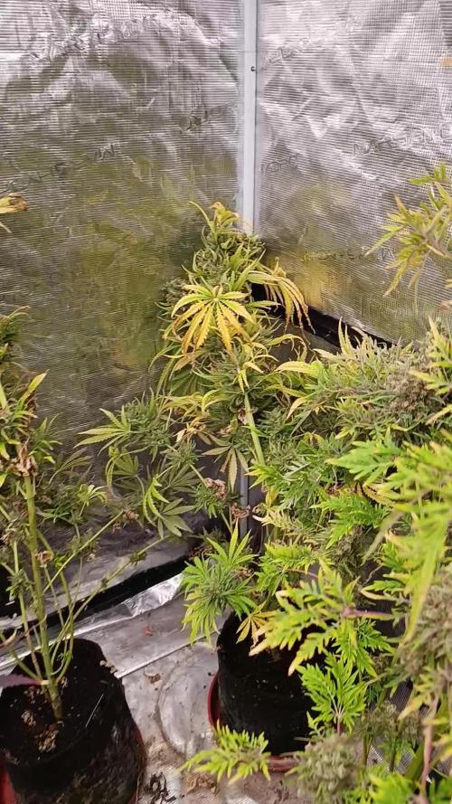
Back 2 are on week 5 day 34 of flowering & front plant Orange sherbet clone is week 8 day 54 & very close to harvest, changed her over & flushing with water & Rezin only. Awaiting delivery of a new 60x microscope lense to view trichomes but the buds look like their just about done. Calyx closed & swollen, amber pistils curling in, they are nice & dense buds.


Likes
19
Share


@adam_pawloski87
Follow
Let’s Go Day 67 from seed !! So this week went real great! 2 Ogreberrys an Bruce Banner started getting flushed! The rest will follow up and start flush next week ! Today the 3 will continue getting flushed while the 3 others get there dose of nutrients, PHd at 6.5! Let’s grow lil ladies let’s grow!!! You all have an amazing productive day as well as a a great safe week !! Peace love and positive vibes to y’all Cheers 😶🌫️💨💨💨💨🤙🏻If there’s any questions please ask, I’ll be opened up to answer at best of my knowledge! Thank you all have a dank day !!

Processing
Likes
12
Share


@blazeblaze
Follow
Soil medium : Coco perlite
I'm running these under 4 6500k 20w LED bulbs and 3 14w 3000k led bulb.
Plant did well the first week.
No nutrients were given.
Stay tuned. stoned*


Likes
8
Share


@Growformyself
Follow
ich habe die Temperatur etwas senken können, die Lampe reduziert und etwas höher gehängt. Mal schauen wie die Woche verläuft.


Likes
15
Share


@Cannibalgardens
Follow
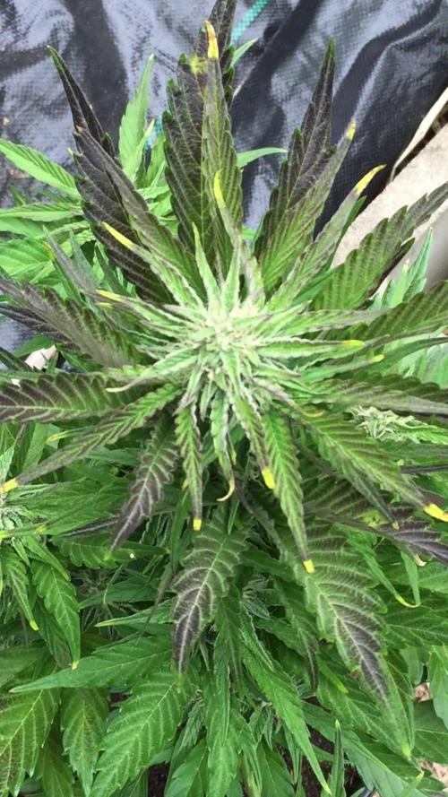
Dam these two have just blown my mind in terms of ability to handle stress and environmental changes . The platinum bananas is the best looking plant so far the healthiest look and vibe , the ripper is also doing very well and looking very bright green and strong smell.. I'm going crazy till next week and can't wait for that flower time ..cheers


Likes
11
Share


@Divs_darkroom
Follow
This girl grew up to be 77 days old. She grew short and without training, focused most of her energy to her main top cola. Even with the cheap Chinese leds shes got dense and really does pack a punch with a strong aroma and flavor. I'm thinking I should probably get more of these beans while they're are still available because she could easily be end up being added to the favorites list. I'll probably put a few nugs away for a long term cure. She was a pleasure to grow.
