The Grow Awards 2026 🏆 


Likes
Comments
Share
@sleeve
Follow
Thanks for having a look at my first photo period grow.
Just starting week 4 of flower. Buds starting to slowly swell, lots of pistils and the leaves are starting to frost.
All lower leaves and branches are removed.
Watering a gallon of bubbled water every second day with my General Organics feed schedule

Likes
50
Share
@Mr_Motalovah
Follow
Hello Growers and Tokers! 👋 👩🌾 🧑🌾.🔥💨
Sorry for the delay on the weekly updates, had some things I had to attend but I'm back and at it!
These pictures were taken a week after the 21 day flower defoliation, day 27 of flower to be exact.
Great progress this week with the bud site, glad to see the defoliation is giving it's fruits.
Buds are filling in space which is just what I want now, fill up as much space as possible that way in the fast weeks they can fatten up and be nice dense colas.
CC1 & CC3 have the best trichome production and bud growth so far despite having the structure all messed up and uneven.
I got some nice pics in of that half loop I was talking about last week. Every time I see it I think, ¨Man, I want to grow a canna bonsai."
CC2 is doing great, bud formation is going a bit slower than the other two but it's still nice.
I'm rooting for this one in highest yield because between the three because she has the most evenly divided canopy.
The Aroma got MUCH stronger this week. When I open up the tent it's a sweet caramel punch to the face.
Don't mind that punch at all! 😍😍
I've had to raise all my plants because the San Fernando Lemon Kush stretched more than I expected.
Bad calculation on my part given it's a sativa I should have expected it would triple in height.
So now I've got all my ladies on top of shoe boxes, Playstation box, old ballast boxes, pretty much any sturdy box I could fix.
Before putting them in there I cleaned them down, making sure nothing unwanted would be joining the garden.
Things look good so far, next week I'll be adding an organic PK boost to the feed. For that extra boom in bloom.
Hope you're enjoying this progress like me.
Have a great day!
One love!

Likes
7
Share
@Robbaus
Follow
Stanno iniziando a gonfiarsi i fiori, anche se devo dire che mi aspettavo qualcosa di più già a questo punto della fioritura, speriamo che esplodano come si deve da adesso in avanti. Le foglie hanno iniziato a diventare gialle, manca poco, credo che in un mese dovrei raccogliere. Ho messo solo due vasi invece che tre come l'ultima volta, ma penso che continuerò così, credo di riuscire a raggiungere lo stesso risultato in termini di resa, e risparmio decisamente sui fertilizzanti. Vedremo

Likes
3
Share


Likes
41
Share
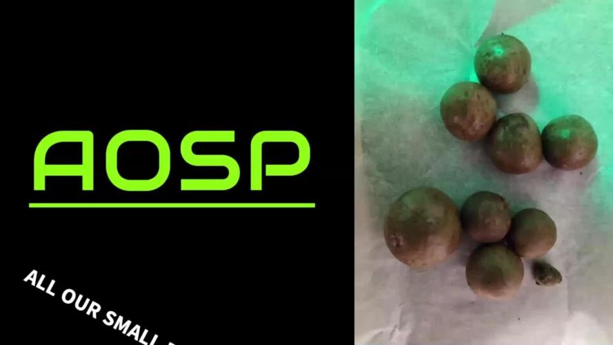
@All_our_small_plants
Follow
Tag 167 heute haben wir nach einer Kontrolle bemerkt das es vermehrt zu Schimmel Bildung kam, und wir haben ein paar Bananen in den Blüten Gefunden, Vermutlich ist es zu viel Stress für die Lady. Mal warm dann wieder kalt, Hohe Luftfeuchtigkeit die ganze zeit. Wir haben nun ca.2,5 KG Nass Gewicht geerntet und werden aus diesem Hash machen, sobald es gefroren ist und wir es verarbeitet haben folgt nochmal ein update hier.
Alles in allem war es eine Interessante Pflanze und wir werden sie nochmal im indoor Testen.
Wir haben nun aus den 2,5 KG Frozen Hash gemacht. Dabei werden frisch geerntete Cannabis-Pflanzen sofort eingefroren und anschließend mit Eis und Wasser verarbeitet, um die Trichome (Harzkristalle) zu lösen und zu Haschisch zu pressen.
Wir haben dazu Bubble bags genommen, diese haben verschieden Micron-Größen: Typischerweise zwischen 25 µm und 220 µm. Je kleiner die Zahl, desto feiner das Sieb und desto reiner das Endprodukt.
Wir haben dazu ein kleines Video hochgeladen wo man den Ganzen Prozess sehen kann.
am ende haben wir dann ein paar schöne Temple Balls erhalten und diese waren einfach genial!!!


Likes
9
Share
@Fullbustero
Follow
This week seems kinda no improvement. I have this feeling but i cant expplain why.


Likes
5
Share
@CreamyNuggets
Follow
Day 64. Harvested the AK -47 and the tallest bubba pupil. The rest of the plants are flushing out and will be harvested around 70 days.

Likes
2
Share
@Reskap
Follow
J’ai fait un peu de lst et de défoliation. J’ai des mouches de terreaux j’ai donc ajouter des pièges collant jaunes. Mes plantes sont tout de même petites quand je compare à d’autres journaux mais c’est la première fois. D’ailleurs si vous avez des conseils pour faire mieux je suis preneur


Likes
Comments
Share
@The710Garden
Follow
Harvest in week 10 with a good amount 50g dry 😜😉
Smooth smoke not a couchlocker a good all day smoke for sure!


Likes
10
Share
@mr_smooke
Follow
flush has started 4 days ago. Her buds are swelling up and she is getting pretty close. Trichomes are mostly cloudy. here is day 56 of flowering


Likes
1
Share
@Skinnytalls420
Follow
#1-03/02/23 the first one to come down!!! Will update as I chop them down!!!

Processing
Likes
16
Share
@Iop420
Follow
Hi all and thanks again for watching my post🤗. From now it's all about waiting and patience😁. Next step is flushing, I think to start next week. I let the pictures and grow log to speak. Peace✌️


Likes
50
Share
@valiotoro
Follow
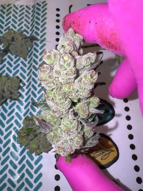
The buds were easy to trim, except they were super sticky but that’s always a good thing! I got two phenos: the first one with that garage-like aroma, and the second one, 100% Blue Cheese!
Processing
Likes
7
Share
@gablmo
Follow
Finally flower. They are 4 feet now. What am I going to do? I don't want them to burn into the light.

Likes
80
Share
@Roberts
Follow
AK 2.0 XL Autoflower has some big saggy buds on her. She grew great through the grow. Minor issues with ph here, and there. I have tried growing with low tds, and high tds in DWC. I prefer High TDS to help control ph level better. Plus plant gets much bigger. From my experiments. This plant is a fine example. Heavy yield, and full of frost. Strong aroma of citrus and a earthy fruity smell. She grew really well under the Mars Hydro FC4800. I will upload a harvest video if it let's me. If not it can be found on my YouTube channel. Link will be below. Thank you Mars Hydro, and Ganja Farmer. 🤜🤛🌱🌱🌱
Thank you grow diaries community for the 👇likes👇, follows, comments, and subscriptions on my YouTube channel👇. ❄️🌱🍻
Happy Growing 🌱🌱🌱
https://youtube.com/channel/UCAhN7yRzWLpcaRHhMIQ7X4g


