
Likes
Comments
Share


@SpartacaZ
Follow
L unica che mi piace sto giro sono la gorilla e la runtz....le piramidi non piacciono...


Likes
19
Share


@Lilside
Follow
Starting to flush out the plant I'm using the flawless finish going to 2ml for ph'd litre of water twice a week and between 1 liter ph'd water I hope this last week of flawless flushed them out
there looking super good and in ready to harvest but I am not sure they all are
I would also like to have them all have a couple days of darkness

Likes
6
Share


@Aleks555
Follow
Zamnesia - Pineapple Express (F1 Auto) - 76 Days
The time has come—harvest day! It's bittersweet to say goodbye to this incredible journey, but all great things must come to an end. The Pineapple Express (F1 Auto) has been a true wonder, delivering both in strength and flavor, with an aroma that's unforgettable. This strain produced the hardest buds we've ever grown—rock-solid and packed with resin, the likes of which we’ve never seen before.
A massive thank you to Zamnesia for providing these remarkable seeds! And an extra special shoutout to Xpert Nutrients for the outstanding fertilizers. The results speak for themselves—your nutrients truly work miracles, creating those dense, powerful buds that we're so proud of.
Stay tuned for the final photo and video showcase of this incredible harvest. We're thrilled to share it with you all!
Xpert Nutrients – The Key to Powerful Harvests
When it comes to achieving the best results, we trust only proven solutions. And with Xpert Nutrients, our plants are getting nothing but the best! These nutrients are not just a product, they’re a true catalyst for growth and powerful yields. Thanks to a balanced formula, each dose nourishes our plants with essential elements, promoting their health, strength, and abundant harvests.
Every cycle with Xpert Nutrients shows progress. We see incredible results: dense, resinous buds, rich aromas, and, of course, yields we can be proud of. With these nutrients, there are no compromises – only maximum performance!
Thank you, Xpert Nutrients, for your continuous support and for helping us unlock the full potential of our plants. The results speak for themselves!

Likes
3
Share
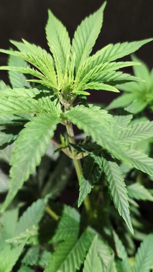

@GoodTimesOrganics
Follow
Hi liebe Community! 💚
Der zweite Strain von Good Times Genetics. Entwickelt sich wunderprächtig und es sind sehr schöne große und Breitgefächerte Blätter zu erkennen. Ein schönes Wuchsbild verreint mit einer starken Indica Dominanz.
Nach dem ausselektieren und dem anwachsen im neuen Topf, ging es letzte Woche noch an dass Toping der Pflanze.
Dieser Genotyp entwickelt sich eher nahe liegend am Hauptstamm, was wir vom Wuchsverhalten aber eher nicht wünschen. Daher wird dieser Typ einfach für den Taste gegrowt.
Wir sind mega gespannt, wie sich die Pflanzen weiter entwickeln.
Nach der nächsten Woche Erholung wird noch ein Topping folgen.
Die Wachstumsbedingungen im Growzelt sind weiterhin am Optimum und Stabil:
———————
🌞 Temp: 23°C
🌚 Temp: 20 °C
💨 RH: 58%
VPD: 0,86 kPa
😎PPFD: 330 mqm
———————
Stay Tuned! 💚
Likes
23
Share


@dataTwiiix
Follow
06/26: très content les fleurs sont givrées à souhait j'ai l'impression qu'un petit coup de pouce minérale ne fait pas de mal à nos chères plantes. #41
06/27: la floraison s'accélère à vue d'oeil :D #42
06/28: arrosage *engraisser
06/29: très content de l'effet des 0.5ml/L de BigBud ajouter à la dernière mixture, prochain arrosage à l'eau ph'd plus additif non NPK.
06/30: une journée de plus au paradis. J'ai augmenter le nombre de lux à 90000 par endroit je verrais demain matin le résultat s'il y en a un.... ms bon max c'est 85000lux j'ai lu ms bon les led dégage moins de chaleur on peut un peu overkill niveau lux le soleil se gêne pas!
07/02: ayant démarrer la culture d'une Orange Sherbet aussi de chez fb42 (je suis conquis par fastbuds42) j'ai donc laissé ce journal pendant deux jours..
Arrosage 3.5L *engraisser (GHE essentiel, BigBud, BudCandy)
Prochaine arrosage avec engrais organique en micro dosage.


Likes
15
Share


@GodG420
Follow
Amazing week for my girls! They are looking lovely! I feed them with 500ml every other day which will be increased to a liter of solution next week. Removing lower fan leaves as usual. Great new is I was chosen from Marshydro and they have sponsored me with sweet new light TS1000! I want to share my thankfulness and happiness!
I will veg my babies in one week and will switch them to 12-12.
Likes
43
Share


@Bones_1986
Follow
Trichomes are 100% cloudy
Plan to flush tomorrow when the pot is dry and harvest Sunday which will be day 68.
Day 67 :
Decided to flush one more time for a final push.

Likes
22
Share


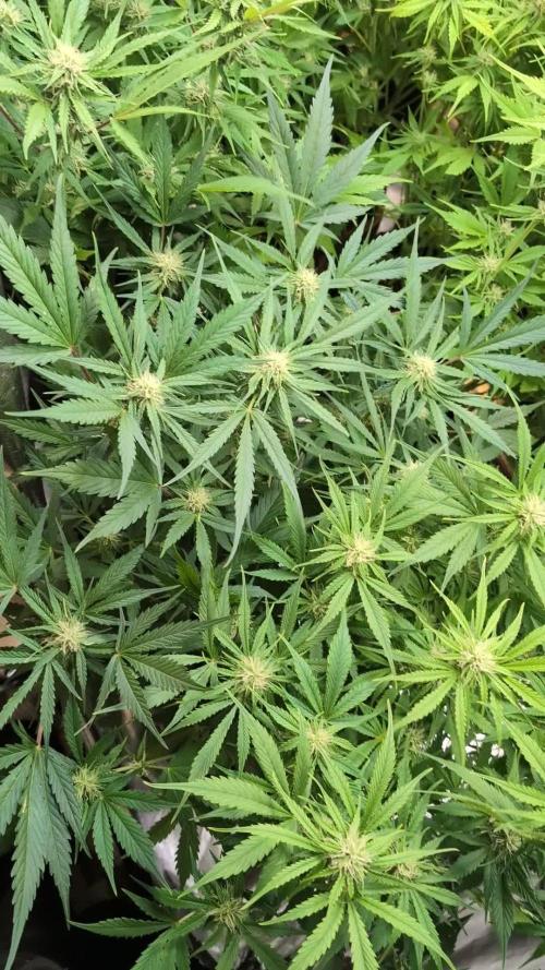
@DeepRootsGrowTrees
Follow
GSC BY KANNABIA
WEEK #18 March 15th-22nd
Week #8 Flower
This week she is looking good. Buds continue to gain weight with a tight bud structure that is covered with trichomes.

Likes
2
Share


@TheHazeMeister420
Follow
Im really hopeful for this round. Not al plants are outside but end of the week they wil be.

Likes
15
Share


@Springbokke
Follow
Доброго времени
Сегодня 82 день жизни моей девочки. Она во всю цвете и сегодня ровна неделя с того дня как я начал считать цвет. Планирую еще 7-8 недель ее продержать, хотя по паспорту у нее всего 8 недель цвета. На 76 день жизни, я обрезал почти все слабые боковые стебли и все нижние листья. За неделю она заметно подросла. Азот я не стал еще убирать, думаю его высокое содержание продержать еще неделю, затем буду уменьшать его содержание в питательном растворе.
Внимательные люди заметили б однозначно весящую лампу, у меня все время вылетало с головы напить о ней))) Эта лампа UVB на 25вт, для рептилий. Много читал про ультрафиолет, единого мнения нет. Это мой второй опыт связанный с ней. Весной этого года я ее использовал с 4 недели цвета, режим работы 4 часа в сутки. Признаюсь я не понял разницу, то есть я не знаю прибавила ли она содержание ТГК в шишках или нет, но точно сказать могу вреда от нее не было. Следующий гров я ее уже не использовал, а в этот раз решил опять воспользоваться ею. На мой взгляд 25вт очень мало, при том что у моих основных ламп 600вт, в будущем думаю купить еще две на 25вт. Если у кого есть опыт с подобными лампамы, пишите, интересно узнать какого результата вы добились.
Полил ( 1212ppm )-3л Sensi Bloom Coco A - 4мл/л; Sensi Bloom Coco B - 4мл/л; Big Bud Coco - 2мл/л; Bud Candy - 2мл/л; Влажность 35% - 47%; температура 21c - 28c 1 - й день цвета, Полил ( 1123ppm )-3л Sensi Bloom Coco A - 4мл/л; Sensi Bloom Coco B - 4мл/л; General Hydroponics Kool Bloom -1мл/л; B-52 - 2мл/л Влажность 32% - 50%; температура 22c - 28c 2 - й день цвета, Полил ( 1081ppm )-3л Sensi Bloom Coco A - 4мл/л; Sensi Bloom Coco B - 4мл/л; Big Bud Coco - 2мл/л; Cal-mag Xtra - 0,5мл/л; Влажность 33% - 50%; температура 22c - 29c
3 - й день цвета, Полил ( 1173ppm )-3л Sensi Bloom Coco A - 4мл/л; Sensi Bloom Coco B - 4мл/л; General Hydroponics Kool Bloom -1мл/л; B-52 - 2мл/л Влажность 31% - 47%; температура 21c - 29c 4 - й день цвета, Полил ( 986ppm )-3л Sensi Bloom Coco A - 4мл/л; Sensi Bloom Coco B - 4мл/л; General Hydroponics Kool Bloom -1мл/л; Bud Candy - 2мл/л; Влажность 32% - 48%; температура 21c - 29c 5 - й день цвета, Полил ( 1121ppm )-3л Sensi Bloom Coco A - 4мл/л; Sensi Bloom Coco B - 4мл/л; General Hydroponics Kool Bloom -1мл/л; Nirvana - 2мл/л; Влажность 30% - 48%; температура 21c - 28c 6 - й день цвета, Полил 3л Влажность 30% - 43%; температура 21c - 27c 7 - й день цвета,

Likes
11
Share


@Chow_13
Follow
Aug 12 - Temp(High/Low) 20/14 Rain(mm) 0.0
Aug 13 - Temp(High/Low) 24/12 Rain(mm) 0.0
Aug 14 - Temp(High/Low) 26/13 Rain(mm) 0.0
Aug 15 - Temp(High/Low) 27/16 Rain(mm) 0.0
Aug 16 - Temp(High/Low) 28/16 Rain(mm) 1.9
Aug 17 - Temp(High/Low) 26/17 Rain(mm) 0.0
Aug 18 - Temp(High/Low) 24/17 Rain(mm) 0.0
Nice week, Buds are looking good. Lots of crystal and white hairs.
Processing
Likes
Comments
Share


@Blind_japanese
Follow
I saw the first pre flowers today. She looks healthy, great growing.
Still struggling to get ph down. Watering with 5.8 ph water.

Likes
18
Share


@Grower_Of_Persia
Follow
my dry and cure style is this:
4 days of hanging upside down to get water activity lower to around 0.6 in 50% humidity and 26 C temp (i know its a little high but we are in a hot summer right now and i cant get it lower even with air conditioner) and then after 4 days of drying i remove leaves and stalks, trim buds and move them to jar for the rest of their life :D . and in the first 4 days of curing i open the jar door and let hem get some fresh air in the jar for about 5 minutes and close the jar door again, after 4 days of curing like that buds are smokable but they will get better as they getting cured about 1 month.
buds are one of the hardest as fucking rocks type of buds! very dense , compact , sticky , smelly , amazing at every aspect
growing stage was 56 days and flowering stage was 75 days total (harvested tops at day 64th)
the total weight of dry buds was : (plant #1 & #3 top buds 56 G + lower buds 22 G ) 78 G + (plant #2 top buds 47 G + lower buds 18 G ) 55 G + (plant #4 top buds 120 G + lower buds 67 G ) 187 G = 367 G

Likes
18
Share


@FrostRailz
Follow
I love the deep green leaves on her , slowly dropping temps to bring out her deep beautiful purple/black, all is well!

Likes
46
Share


@exclusiveX420
Follow
Amazing week, my first ever harvest for the 1 gallon gorilla glue auto. I was afraid to wait any longer as she had nearly no leaves left. She did have alot of milky Trichomes but still some clear around, few amber. Currently drying and awaiting results. I was unable to get an accurate wet wait as I left the stalk and all. I tried my best and saw 126g go across the scale but I just layed it quick before it fell over. I'm hoping 1/2 ounce usable yield when dry as it was not nearly as dense as I thought. The other plants are much more dense and continue to harder so I will be waiting as long as they need. They are very healthy. Praying they finish with high quality. 🙏 GSCE is VERY dense feeling though and GG 2 gal is catching up but not super solid yet. BB still taking her good ole time. Maybe within a month ? I sure hope because I'm running out of time for this project until summer. Praying for a fast , solid finish! 🙏 Less than 2 weeks for sure for GG & GSCE , they are extremely covered and mostly milky just some clear still and not enough amber. I have lowered the light an inch every day or so for the last few days. I'm now breaching the limit but still a hair of room. Hoping I can achieve the 10-30% amber and have something special. Stay tuned

Likes
2
Share


@Organic473
Follow
Just been feeding her water once a week..she continues to swell I will give her some molasses one for the road and just water until week 8.
Likes
5
Share

@smoking_hills9
Follow
Very happy I went Original Sensible Seeds beens this grow. 🤩 All 3 autos from some special american line, and o boy , they don't disappoint.🔥 Bangers genetics 👏🏻 all trees are good size with super thick stalks and loads of fat buds. Another big defoliation been done this week, removed around 2 kg of leaves.All frosty with great smell. I like it a lot 💚

Likes
5
Share


@Tacofever88
Follow
A quick check up and they're doing well. Some growth so that's a good sign.
Some little bastard animal (mole I assume) made a few holes around my girls. I made sure to put some fluid in them as I was peeing. Then stuffed them with rocks, dirt and a thick branch. Hopefully they're not too interested and I don't have an animal having a root party
Processing
Likes
41
Share


@Hawkbo
Follow
Day 42, everything is chuggin along pretty healthy. Definitely some seeds in most of them which bugs me. The whole tent reeks tho which is nice. Some of the citradellics look like they are gettin pretty close so I'll start keeping an eye on them. I had to take the pics again and was strugglin so they arent my best. I have to go thru them still so I got the video up and tmm or as soon as possible I'll get the still shots up✌️
2 and 3 look pretty good but 1 is on another level right now shes sweatin like a hoe in church. The aromas coming off her take over the room like rotten citrus it's one of my favorite smells. 1 and 2 have more awkward looking bud structures but still decent. 1 was so under wated before last nights feed it was flopping over everywhere nearly dead but after an hour showed improvement, it just dries out so much quicker than everything else it's hard to keep up with.

Likes
4
Share


@Slobasian
Follow
Sunday last day of week 11 expecting to cut it down fed banana, molasses, and honey for a final flush with ice, Friday final ice flush. In the dark for 12 hours then cut down for harvest