
"Jack Herer" C#4


VEG
500w Light Emitting Diodes/600W
500w


Bone Meal Other
Blood Meal Other
Fish Bone Meal Other
Kelp Meal Other
Gypsum Other
Wollastonite Other
Dolomitic Lime Other
Basalt Rock Dust Other
Humic Acid Other
Mycorrhizae Other
Worm Castings Other
Azomite Other
Greensand Other
Shrimp Chitin Other

Perlite

Vermiculite

Coco Coir
Supercropping
weeks 17
LST
weeks 8
Main-Lining
weeks 8
Defoliation
weeks 8
100 l
Pot Size
Start at Harvest
G
Germination4y ago


Nutrients 1
Coconut water
100 mll
Ultraviolet ultra-
7 likes
3 comments
Share
Used method
Other
Germination Method
8
Week 8. Vegetation4y ago


45.72 cm
Height
16 hrs
Light Schedule
30 °C
Day Air Temp
6.0
pH
300 PPM
TDS
45 %
Air Humidity
25 °C
Solution Temp
22 °C
Substrate Temp
14 °C
Night Air Temp
0.98 l
Pot Size
0.49 l
Watering Volume
45.72 cm
Lamp Distance
1250 PPM
CO₂ Level
Ultraviolet nopeeee
5 likes
comments
Share
Used techniques
LST
Technique
Main-Lining
Technique
Defoliation
Technique
9
Week 9. Vegetation4y ago

45.72 cm
Height
16 hrs
Light Schedule
29 °C
Day Air Temp
6.0
pH
300 PPM
TDS
45 %
Air Humidity
25 °C
Solution Temp
22 °C
Substrate Temp
18 °C
Night Air Temp
0.98 l
Pot Size
0.49 l
Watering Volume
45.72 cm
Lamp Distance
1250 PPM
CO₂ Level
Ultraviolet Leaf shape and size are determined by the level of light captured within the leaf. Once the danger of heat radiation burning the leaves has been negated, there is no such thing as too much light; there is a point of diminishing returns in terms of photosynthesis, but so long as you keep temperatures under control, "too much light" is not a thing. The plant uses its photoreceptors to keep accurate tabs on light levels around the plant, it uses this feedback that determines where it puts its resources moving forward. In low light and low temperatures, the plant will produce big fat thick leaves. This is by design as there are few photons available, so it must make sure it captures as many photons as it can within the chlorophyll net inside the leaf epidermis, hence It makes it as thick and dense as it can. Once the plant reaches high levels of light saturation and toasty temperatures, you will noticeably see much smaller and thinner leaves form as the plant gets closer to high-level light sources. Useful to a knowing eye, you can ballpark estimate if a light source is too far away just from looking.
Plant life is hardwired to grow toward the light. With the introduction of LEDs, heat radiation is no longer a big issue. Left to its own intelligence, a plant will slow down vertical growth once it can no longer grow vertically comfortably. By topping once and then some lst bend a (highest) main stem below any other plant will now devote all its resources to whichever stem/s are nearest the light source. Just keep bending them all over, and it will seem like the plants grind to a halt, distributing their little resources to so many main stems.
5 likes
comments
Share
11
Week 11. Vegetation4y ago


55.88 cm
Height
16 hrs
Light Schedule
27 °C
Day Air Temp
6.5
pH
300 PPM
TDS
45 %
Air Humidity
25 °C
Solution Temp
21 °C
Substrate Temp
14 °C
Night Air Temp
0.98 l
Pot Size
0.49 l
Watering Volume
45.72 cm
Lamp Distance
1250 PPM
CO₂ Level
Ultraviolet A cutting requires roots to uptake water, we must create an environment so humid the plant can absorb moisture through its one set of leaves, 70+RH% but also a good supply of fresh air and full temperature control. Once I see light lime green it will signify new growth of the plant, I will know it has started to re-root.
5 likes
1 comment
Share
13
Week 13. Vegetation4y ago


101.6 cm
Height
24 hrs
Light Schedule
27 °C
Day Air Temp
6.5
pH
300 PPM
TDS
45 %
Air Humidity
25 °C
Solution Temp
21 °C
Substrate Temp
14 °C
Night Air Temp
0.98 l
Pot Size
0.49 l
Watering Volume
45.72 cm
Lamp Distance
1250 PPM
CO₂ Level
Ultraviolet Switched to 12 hours of dark. Just waiting the 4-7 days for first sign of flowers to show before I can start counting the flowering weeks.
I want to take cuttings for clones when stem is juiced with flowering hormones.
6 likes
comments
Share
14
Week 14. Flowering4y ago


101.6 cm
Height
12 hrs
Light Schedule
27 °C
Day Air Temp
6.5
pH
300 PPM
TDS
45 %
Air Humidity
25 °C
Solution Temp
21 °C
Substrate Temp
14 °C
Night Air Temp
3.79 l
Pot Size
3.79 l
Watering Volume
45.72 cm
Lamp Distance
1250 PPM
CO₂ Level
Ultraviolet aw well
6 likes
comments
Share
15
Week 15. Flowering4y ago


101.6 cm
Height
12 hrs
Light Schedule
27 °C
Day Air Temp
6.5
pH
300 PPM
TDS
45 %
Air Humidity
25 °C
Solution Temp
21 °C
Substrate Temp
14 °C
Night Air Temp
3.79 l
Pot Size
3.79 l
Watering Volume
45.72 cm
Lamp Distance
1250 PPM
CO₂ Level
Nutrients 2
Aluminum Sulphate
100 mll
Love
50 mll
Ultraviolet Light is an important environmental factor which controls growth and development in plants. Besides photosynthesis in which light is harvested by green plants and is converted- into chemical energy, there are numerous other plant responses to light such as phototropism, germination of some light sensitive seeds e.g. lettuce, de-etiolation of monocot and dicot seedlings etc., which are quite independent of photosynthesis and in which light just acts as environmental signal to bring about the particular photo-response.
Most of these photo-responses control genetically defined structural development or morphogenesis (i.e., origin of form) of plants. The role of light in regulating morphogenesis is known as photo-morphogenesis. In plants, red and blue light are especially effective in inducing a photo-morphogenetic response.
The effect of light in controlling morphogenesis can best be demonstrated by comparing a monocot (maize) or dicot (bean) seedling grown in light with one grown in darkness both of which have been reared from genetically identical seeds. Abundant reserve food in seeds eliminate the need for photosynthesis for many days.
It can easily be noticed that dark grown seedling has become etiolated (i.e., pale and weak) while the one grown in light has stockier and green appearance with short stem and large leaf area (fig1. Since both etiolated and light grown seedlings were reared from genetically identical seeds, light must have altered the gene expression during germination so that the appearance or form of etiolated and light grown seedlings looks different.
De-etiolation of light grown seedling can be done in very short period (hours) by placing it even in dim light. During de-etiolation, marked reduction in the rate of stem elongation, straightening of apical hook and development of green pigments can easily be noticed. The etiolated form of the seedling is thus gradually transformed to stockier green appearance and is the result of photo-morphogenesis. The development of seedling in darkness is called as skoto-morphogenesis (from Greek word Skotos = darkness).
According to Hans Mohr (1983), there are two important stages of photo-morphogenesis:
(i) Pattern specification, in which cells and tissues develop specific ability or competence to respond to light during certain developmental stage and
(ii) Pattern realization, during which time the photo-response occurs.
There are two main categories of plant responses to light signals:
(i) Phytochrome mediated photoresponses and
(ii) Blue-light responses or cryptochrome mediated photo-responses.
(A) Phytochrome Mediated Photoresponses in Plants:
Large number of photo-morphogenic responses in plants are known to be mediated by the proteinaceons pigment (chromoprotein) called phytochrome. This pigment acts as photoreceptor and absorbs most strongly red and far-red light. It also absorbs blue light.
The pigment phytochrome exists in two forms, (i) a red light absorbing form designated as PR form and (ii) another far-red light absorbing form designated as PFR form. These two forms are photochemically interconvertible. When PR form absorbs red light (650 – 680 nm), it is converted to PFR form. The PFR form absorbs far-red light (710 – 740 nm) and is converted to PR form. The PFR form of this pigment is believed to be physiologically active form.
Absorption spectra of PR and PFR forms of phytochrome purified from etiolated Avena seedlings are given in Fig. 25.2. PR form shows a peak at 666 nm while PFR form at 730 nm. It is noteworthy that both forms of phytochrome also absorb in the blue region of spectrum of light.
(The absorption maxima obtained in vitro mostly correspond with those in vivo provided that the native phytochrome is carefully purified and non-degraded).
However, it is quite obvious from this figure that the absorption maxima of PR and PFR forms overlap considerably in the red region of the spectrum of visible light and therefore, the phytochrome system cannot be quantitatively converted from PR to PFR. After irradiation with red light (or white light), there is an equilibrium between PR and PFR forms that depends upon the spectral composition of the light source. This equilibrium is called as photo-stationary equilibrium (ф) and is defined as ratio of the PFR conc. and the total phytochrome conc. (ρtotal) at a given wavelength. These values can be measured by difference spectroscopy.
ф = PFR/ PR+ PFR = PFR/Ptotal
Most of the phytochrome mediated photoresponses in plants are reversible. These are induced by red light and reversed by far red light. A list of some of the photoreversible responses mediated by phytochrome in plants is given in table 25.1.
Photoreversible responses mediated by phytochrome in plants
One of the classical examples of photo-morphogenesis in plants induced by short red-far- red pulses is the germination of light sensitive seeds of lettuce (Lactuca sativa). In early 1930s, Flint and McAlister (1937) demonstrated that germination of lettuce seeds is not only stimulated by white light but also by red light (shorter than 700 nm) and inhibited by far-red light (greater than 700 nm).
In 1950s, Borthwick and Hendricks and their associates obtained spectacular results by exposing lettuce seeds to alternating red and far-red treatments. They observed much higher percentage of germination when the seeds received red light as the final treatment. Seed germination was markedly inhibited when seeds received final treatment with far-red light. (Table 25.2)
Effect of alternating red/far
Borthwick and his associates also predicted existence of the photoreceptor phytochrome in two different forms which was proved to be absolutely correct later on when this pigment was isolated in plant extracts for the first time by Butler et al in 1959 and its photo-reversibility was confirmed in vitro.
Based on the amount of light required or the fluence (no. of photons absorbed per unit surface area), the phytochrome mediated photo-responses can be grouped into three main categories:
(a) Very Low Fluence Responses (VLFRs):
These responses are initiated by very low fluences (0.1 to 1 n mol m-2) saturating at 50 n mol m-2 and are non-photo reversible. For example, brief flash of red light with fluence as low as 0.1 n mol m-2 can stimulate the growth of coleoptile and inhibit growth of mesocotyl in oat seedlings that have been grown in dark. Similarly, red light with fluence of only 1-100 n mol m-2 is enough to stimulate seed germination in Arabidopsis. (In monocots, the elongated area of axis between coleoptile and root is called as mesocotyl)
(b) Low Fluence Responses (LFRs):
These responses require fluence of at least 1.0 nmol m-2 saturating at 1000 n mol m-2 and are photo-reversible. Most of the red/far-red photo-responses including the lettuce seed germination belong to this category.
(c) High Irradiance Responses (HIRs):
These responses require continuous or prolonged exposure to light of relatively high irradiance saturating at much higher fluences (at least 100 times more) than LFRs and are non- photo-reversible.
Examples are:
(i) Anthocyanin synthesis in dicot seedlings and in apple skin,
(ii) Ethylene production in sorghum,
(iii) Induction of flowering in Hyoscyamus (a long day plant),
(iv) Opening of plumular hook in lettuce,
(v) Enlargement of cotyledons in mustard,
(vi) Inhibition of hypocotyl elongation in many dicot seedlings etc.
(B) Blue Light Responses or Cryptochrome Mediated Photoresponses:
Apart from phytochrome mediated photo-responses, large number of photo-responses in plants are known which are controlled by blue light and are believed to be mediated through a group of yet unidentified pigments called crypto chrome (crypto from cryptogams), the latter acting as photoreceptor in such responses. Blue light responses have been reported in algae, fungi, ferns and higher plants.
Some of the typical and most commonly known blue-light responses in plants are:
(i) Phototropism
(ii) Stomatal opening
(iii) Inhibition of hypocotyl elongation
(iv) Sun tracking by leaves
(v) Phototaxis
(vi) Movements of chloroplasts within the cells and
(vii) Stimulation of synthesis of carotenoids and chlorophylls etc.
Crypto chrome absorbs light rays mostly in violet-blue region of the spectrum (400 – 500 nm). It also absorbs long wave ultraviolet rays in UV-A region (320 to 400 nm). However, most photo-responses of plants caused by crypto chrome result from absorption in violet-blue region of the spectrum but they are simply called as blue-light responses
Although phytochrome and some other photoreceptors also absorb blue light, but the typical blue-light morphogenetic responses differ from photo-responses mediated by them in being insensitive to red light and there is no red/far- red reversibility.
i. The action spectra of many blue-light responses in higher plants such as phototropism, stomatal movement, inhibition of hypocotyl elongation etc. are similar and characteristic. They show three peaks in blue region (400 – 500 nm) of the spectrum of visible light. This three peaked, action spectrum is also known as three fingers action spectrum (because of its resemblance in shape with three fingers) and is typical of most blue light responses (Fig. 25.3). Three fingers action spectrum is not observed in phytochrome mediated photo-responses or photo-responses mediated by other photoreceptors other than crypto-chrome.
Typical three peaked
ii. Scientists have implicated roles of yellow pigment carotenoids or flavins as photoreceptors in blue-light responses of plants for a long time. However, the spectroscopy of blue-light responses is complex and it is not easy to distinguish between these two types of pigments by comparing available action and absorption spectra.
Action spectrum for phototropism
Fig. 25.4. Shows relationships between action spectrum for phototropism and absorption spectra of riboflavin and β-carotene. The strong peak in UV-region of the spectrum (360-380 nm) suggests riboflavin as the photoreceptor pigment, while three peaks in blue regions (400 – 500 nm) of the spectrum favours carotene. Nevertheless, accumulating evidences strongly favour flavin pigment to be the primary photoreceptor in phototropism.
Schmidt (1984) has summarised arguments in favour of flavins or carotenoids as photoreceptor pigments in blue light responses of plants as follows:
(a) Arguments in Favour of Flavins:
(i) Action spectra show UV maximum between 350-400 nm.
(ii) Primary steps of the blue light response are dependent on presence of O2.
(iii) Flavin reactions are often redox reactions.
(iv) Light can be substituted by oxidants while reductants suppress the blue light reaction.
(v) Blue light reaction is inhibited by flavin inhibitors such as KI.
(vi) Blue light action spectra resemble low temp, spectra of flavins.
(vii) Neurospora mutant which is free of carotenoids shows blue light response.
(viii) Half life of carotenoids in first excited singlet state is very short (10-13 seconds)
(b) Arguments in Favour of Carotenoids:
(i) Three peaked (three fingers) action spectra resemble absorption spectra of carotenoids.
(ii) Small or no UV maximum in some action spectra.
(iii) Energy transfer from UV absorbing pigment to carotenoids is feasible.
(iv) Carotenoids from diatom mutant do not show blue light response.
Earlier evidences suggested crypto chrome to be one or both of the yellow pigments, carotenoids (such as β-carotene, zeaxanthin) and/or flavins (such as riboflavin, FAD) which mediate blue-light responses in plants.
However, with extensive researches done with mutants and transgenic plants and over expression studies beginning in early 1990s, the vexed problem of identification of blue-light receptors in plants has gradually been resolved now.
The term crypto chrome is now applied specifically to flavoprotein photoreceptor that mediates inhibition of hypocotyl (stem) elongation caused by blue-light. Blue-light photoreceptor in phototropism and chloroplasts movements in plants is phototropin which is also a flavoprotein. The carotenoid zeaxanthin is blue-light photoreceptor involved in stomatal opening.
Photoreceptors:
A brief account of all these photoreceptors follows:
1. Crypto-chrome:
The first protein with characteristics of blue-light receptor was isolated in 1993 from Arabidopsis. It was found that hy4 mutant of Arabidopsis had lost the capacity to respond specifically to blue-light in that it showed an elongated hypocotyl even on irradiation with blue-light (In the wild type, blue-light causes inhibition of hypocotyl elongation).
Isolation of the hy4 gene (later named as cryl) showed that it encoded a 75 kDa protein called crypto-chrome 1 (CRY1) with remarkable sequence similarity (homology) to DNA photolyase in having two chromophores: a flavin adenine dinucleotide (FAD) and a pterin attached to the apoprotein (Fig. 25.5). This led to the establishment of cryptochrome to be a flavoprotein that was involved in inhibition of hypocotyl elongation in response to blue-light. (The structure of pterin is given in figure 25.6. For structure of FAD).
Diagrammatic representation of the chromophere binding domains
(DNA photolyase is a blue-light activated flavoenzyme which repairs UV-induced damage to microbial DNA. Cryptochrome differs from photolyase mainly in two respects. Firstly, the cryptochrome does not show photolyase activity and secondly, unlike photolyase it has an extended carboxy-terminal domain (Fig. 25.5) with kinase activity).
A second cryptochrome 2 (CRY2) also with two chromophores like CRY1, has also been isolated from Arabidopsis (Lin 2000). CRY2, mediates blue-light stimulated inhibition of hypocotyl elongation, increase in cotyledon expansion and anthocyanin production. It also has a role in determining flowering time. Both CRY1 and CRY2 appear to be ubiquitous in plant kingdom, but while CRY1 is stable in light grown seedling, CRY2 is rapidly degraded in light.
Structure of pterin
Mechanism of action of cryptochrome:
The mechanism of action of crypto chrome remains elusive so far. The flavins are known to participate in oxidation-reduction reactions and photolyases repair damaged DNA (as a result of UV-radiations) by transferring electrons to pyrimidine dimers. Crypto chromes may act probably in a similar way through some electron transfer mechanism.
2. Phototropins:
Phototropins are blue-light receptors that mediate phototropism and chloroplasts movements in plants. In late 1980s, it was found that blue-light stimulated phosphorylation of a 120 kDa protein located on plasmamembrane of actively growing regions of etiolated seedlings. These regions were also most responsive to phototropic stimulus. Extensive biochemical and physiological studies showed this protein to be a kinase autophosphorylating in blue-light and which could be the photoreceptor for phototropism.
Later on, a mutant nph1 (won phototropic hypocotyl 1) was isolated from Arabidopsis which lacked phototropic response in the hypocotyl and also the 120 kDa membrane protein. It was genetically independent of the hy4 mutant as it showed blue-light induced inhibition of hypocotyl elongation.
The nph1 gene was cloned and it was found (as postulated) to encode a 120 kDa protein nph1. The nph1 gene was renamed as phot1 and the protein encoded by it was named phototropin (Briggs and Christie, 2002).
Phototropin is also a flavoprotein with two flavin mononucleotide (FMN) chromophores. The protein has a carboxy-terminal domain with a serine/threonine kinase activity. In the amino-terminal half, there are two domains called LOV domains (of about 100 amino acids each) to which are attached the chromophores (Fig. 25.5). (LOV domains are so called because they are characteristics of microbial proteins which regulate response to light, oxygen and voltage).
Recent spectroscopic studies done by Swartz et al, 2000) have shown that in dark, FMN molecules remain non covalently bound to LOV domains, but on irradiations with blue-light they become covalently bound to cysteine residues of the apoprotein through a sulphur atom forming a cysteine- flavin covalent adduct. The reaction is reversed in dark. A second gene called phot 2 has also been isolated from Arabidopsis which is related to phot 1. It is believed that phototropic response involves both phot 1 and phot 2.
Mechanism of action of phototropins:
The mechanism of action of phototropins is not clear. It has been observed that blue-light causes a transient increase in cytosolic calcium concentration and there are indications that phototropin signalling chain may partly involve regulation of cytoplasmic calcium concentration.
3. Zeaxanthin:
The carotenoid zeaxanthin has been shown to be blue-light receptor in guard cells that plays central role in blue-light stimulated stomatal opening. (See chapter 17 for structure of zeaxanthin).
Following evidences strongly support role of zeaxanthin in stomatal opening:
(i) The absorption spectrum of zeaxanthin closely resembles the action spectrum of blue- light stimulated stomatal opening.
(ii) During stomatal opening in intact leaves, the incident radiation, zeaxanthin concentration in guard cell, and stomatal apertures have been found to be directly correlated.
(iii) Blue-light sensitivity of guard cells increases with an increased concentration of zeaxanthin in guard cells.
(iv) There is complete inhibition of blue-light stimulates stomatal opening by 3mM conc. of dithiothreitol (DTT) which is a potent inhibitor of the enzyme that converts violaxanthin to zeaxanthin.
(v) In facultative CAM plant species such as MeSembryanthemum crystallinum, there is a shift from C3 to CAM mode of carbon metabolism in response to accumulation of salts. In C3 mode, the guard cells accumulate zeaxanthin and exhibit blue-light response. But, in CAM mode, neither there is accumulation of zeaxanthin in guard cells nor they respond to blue- light. (In CAM plants, stomata remain closed during the day).
Mechanism of action of zeaxanthin:
It is believed that the excitation of zeaxanthin by blue- light in guard cells starts a signal transduction pathway that includes:
(i) Isomerization of zeaxanthin,
(ii) Conformational changes in the apoprotein
(iii) Transmission of blue-light signal across the chloroplast membrane by a secondary messenger (most probably Ca++, phosphatases, calcium binding protein calmodulin and inositol triphosphate (IP3),
(iv) Activation of H+-ATPases at the guard cell plasma membrane resulting in pumping of protons across the membrane and intake of K+ ions followed by Cl– ions.
(v) Turgor build up in guard cell and stomatal opening.
The blue-light stimulated stomatal opening can be reversed by green light. This may happen if green light is applied with blue-light in continuous light treatment or if a blue-light pulse (of about 30 seconds duration) is followed by a green light pulse. A second blue-light pulse after green-light can restore the stomatal opening. It has been suggested by various workers that green light reverses the isomerization of zeaxanthin resulting in regeneration of inactive zeaxanthin isomer. The latter is unable to mediate the blue-light response.
(Besides phytochrome and cryptochrome, there are two other categories of photoreceptors which are known to affect photomorphogenesis in plants. They are, (i) protochlorophyllide-a, a pigment which absorbs red and blue light and is converted to chlorophyll-a and (ii) UV-B photoreceptor – one or more unidentified compounds which absorb short wave ultraviolet rays in UV-B-region (280-320 nm).
7 likes
comments
Share
16
Week 16. Flowering4y ago


101.6 cm
Height
12 hrs
Light Schedule
27 °C
Day Air Temp
6.5
pH
300 PPM
TDS
45 %
Air Humidity
25 °C
Solution Temp
21 °C
Substrate Temp
16 °C
Night Air Temp
26.5 l
Pot Size
3.79 l
Watering Volume
45.72 cm
Lamp Distance
1250 PPM
CO₂ Level
Nutrients 2
Aluminum Sulphate
100 mll
H202 3%
100 mll
Ultraviolet Hydrogen peroxide (H2O2), occurs naturally in rainwater and acts as nature's cleanser. It oxygenates soil and bodies of water, is completely non-toxic and safe to use around food, people and animals. Adding it when watering your indoor plants, mimics what Mother Nature does for outdoor plants every time it rains.
Hydrogen peroxide is distilled water with an extra atom of oxygen. H2O means 2 hydrogens and 1 oxygen atom. The extra 2 in H2O2 means 2 hydrogens and 2 oxygen atoms, called hydrogen peroxide.
H202 rapidly decays in the soil decaying Into h2o and o.
Oxygen is the number ONE limiting factor of plant growth (once supplied with co2).
The Oxygen is mixed with water, starches sugars, n,p and k to make the more complex proteins.
H2O2 provides extra oxygen to roots for growth and germination:
To sanitise seeds: Use undiluted H2O2 3%. Put seeds in a container that can be sealed. Pour Oxygen Plus to just cover seeds. Seal and soak seeds for 4 hours. Rinse with water.
To speed up germination: Mix 30mls H2O2 3% with 2 cups of water (a measuring cup makes it easy to get the right amount). Soak seeds overnight, then plant as usual.
For planted seedlings: Spray with H2O2 solution of 1 part H2O2 3% to 32 parts water (eg: around 30mls per 1 litre - measuring cup recommended).
For established plants: Mix 1:1 ratio of 3% H2O2 and water (eg: 1 cup of each). Bottom or top water as usual (water soil only, do not pour on leaves).
5 likes
1 comment
Share
17
Week 17. Flowering4y ago


101.6 cm
Height
12 hrs
Light Schedule
27 °C
Day Air Temp
6.5
pH
300 PPM
TDS
45 %
Air Humidity
25 °C
Solution Temp
21 °C
Substrate Temp
16 °C
Night Air Temp
26.5 l
Pot Size
3.79 l
Watering Volume
45.72 cm
Lamp Distance
1250 PPM
CO₂ Level
Nutrients 1
H202 3%
5.21 mll
Ultraviolet I have been hesitant with super-cropping never fully understanding exactly what to do, worrying, decided to try a few side-stems and it worked very well with little to no damage on the exterior lining, excited to add it to the arsenal of techniques more frequently next grow as it was really noticed the boost in growth on the tested stem.
6 likes
comments
Share
Used techniques
Supercropping
Technique
18
Week 18. Flowering4y ago


101.6 cm
Height
12 hrs
Light Schedule
24 °C
Day Air Temp
6.5
pH
1000 PPM
TDS
45 %
Air Humidity
25 °C
Solution Temp
24 °C
Substrate Temp
18 °C
Night Air Temp
26.5 l
Pot Size
3.79 l
Watering Volume
45.72 cm
Lamp Distance
1250 PPM
CO₂ Level
Nutrients 8
H202 3%
5.21 mll
Aluminum Sulphate
0.65 mll

RAW Potassium
1.3 mll
Ultraviolet ggggg
8 likes
4 comments
Share
19
Week 19. Flowering4y ago


101.6 cm
Height
12 hrs
Light Schedule
24 °C
Day Air Temp
6.5
pH
1000 PPM
TDS
45 %
Air Humidity
25 °C
Solution Temp
24 °C
Substrate Temp
18 °C
Night Air Temp
26.5 l
Pot Size
3.79 l
Watering Volume
45.72 cm
Lamp Distance
1250 PPM
CO₂ Level
Nutrients 2
H202 3%
5.21 mll
Azomite
5.21 mll
Ultraviolet Human Body
65% Oxygen (in all liquids and tissues, bones, and proteins)
18% Carbon (everywhere)
10% Hydrogen (in all liquids and tissues, bones, proteins
3% Nitrogen (in all liquids and tissues, proteins
1,5% Calcium (lungs, kidney, liver, thyroid, brain, muscles, heart, bones)
1% Phosphorus (urine, bones, DNA)
0,35% Potassium (enzymes)
0,25% Sulphur (proteins)
0,15% Sodium (in all liquids and tissues) (in terms of salt)
0,05% Magnesium (lungs, kidney, liver, thyroid, brain, muscles, heart)
The average adult male contains about 140 g of K(Potassium); the level varies with body weight and muscle mass. We ingest about 2.5 g per day of K from our food and excrete about the same amount. 0.0118 % of that is K40
The answer is that they were present when our earth was formed. Any radioactive material originally present at the formation of the earth would have decayed and disappeared if its half-life was short compared to the age of the earth. However, if its half-life were long, close to or greater than the age of the earth, then such materials would not have disappeared but are still with us today.
There are several radioelements in this category, such as the well-known elements uranium and thorium. Thorium (Th232) has a half-life of 14,000,000,000 years, uranium has two long-lived radioisotopes; U238 has a half-life of 4,500,000,000 years, and U235 has a half-life of 710,000,000 years. These give rise to the radium and thorium atoms found in all humans, acquired from the food we eat. That food, of course, obtained these materials from the soil in which it grew or on which it grazed.
Potassium is also in this category. There are actually three potassium isotopes: K39, a stable isotope, is the most abundant, at 93.26 % of the total; K41 is next in abundance at 6.73 % and is also a stable isotope. The potassium isotope of interest is a radioactive isotope, K40. It is present in all potassium at a very low concentration, 0.0118 %. It has a very long half-life, 1,260,000,000 years. When it decays 89 % of the events give rise to the emission of a beta ray with maximum energy of 1.33 MeV. The other 11 % of the decays produce a gamma-ray with an energy of 1.46 MeV
The forces required to forge thorium 232 can only be harnessed when traveling close to or at the speed of light, so essentially what I'm getting at is 0.0118% of every person alive is formed of the same element that was forged in the anvil of creation itself. We are all one & the same
German chemist Johann Wolfgang Dobereiner attempted to classify elements with similar properties into groups of three elements each. These groups were called ‘triads’. Dobereiner suggested that in these triads, the atomic mass of the element in the middle would be more or less equal to the mean of the atomic masses of the other two elements in the triad.
An example of such a triad would be one containing lithium, sodium, and potassium. The atomic mass of lithium 6.94 and that of potassium is 39.10. The element in the middle of this triad, sodium, has an atomic mass of 22.99 which is more or less equal to the mean of the atomic masses of lithium and potassium (which is 23.02). 9 controls the 6 and 3.
The Limitations of Dobereiner’s Triads are :
All the elements known at that time couldn’t be classified into triads.
Only four triads were mentioned – (Li,Na,K ), (Ca,Sr,Ba) , (Cl,Br,I) , (S,Se,Te).
2. Newland’s Octaves
English scientist John Newlands arranged the 56 known elements in increasing order of atomic mass in the year 1866. He observed a trend wherein every eighth element exhibited properties similar to the first.
Azomite contains 180ppm of thorium.
Your plant will thank you, you are welcome.
Most farmers do have not a proper understanding of what is Azomite and how to use it in gardening, especially if they practice organic farming. Continuous propagation and leaching effects of water deplete the essential minerals and micro-nutrients from the soils. Such soils remain weak, not able to support the production of fruits and vegetables. Azomite mineral contains micronutrients that supplement the soil. It also balances the minerals for growth and overall productivity. Constant use of this mineral rejuvenates your soil renewing its potency again. Azomite is a naturally mined mineral product that is ready to use. It’s a unique rock that comes from a mine in central Utah. Azomite requires no mixing or special preparation before use. It is derived from volcano ash that spewed out millions of years ago. It contains the widest range of minerals of all the rock dust in the world. Azomite provides plants with 70% essential elements. These elements include magnesium, calcium, potassium, and silicon for plant growth.
Facts About Azomite Fertilizer
It’s a natural mineral – 100% natural with no fillers or additives
Does not contain any harmful elements
Requires no special preparation before use
It’s odorless – very friendly to use
Does not restrict water penetration or aeration
Is easily broken down and absorbed into the soil
Does not burn plants.
7 likes
2 comments
Share
20
Week 20. Flowering4y ago


101.6 cm
Height
12 hrs
Light Schedule
24 °C
Day Air Temp
6.5
pH
1000 PPM
TDS
45 %
Air Humidity
25 °C
Solution Temp
24 °C
Substrate Temp
18 °C
Night Air Temp
26.5 l
Pot Size
3.79 l
Watering Volume
45.72 cm
Lamp Distance
1250 PPM
CO₂ Level
Ultraviolet gggg
6 likes
1 comment
Share
21
Week 21. Flowering4y ago


101.6 cm
Height
12 hrs
Light Schedule
24 °C
Day Air Temp
6.5
pH
Normal
Smell
1000 PPM
TDS
45 %
Air Humidity
25 °C
Solution Temp
24 °C
Substrate Temp
21 °C
Night Air Temp
26.5 l
Pot Size
3.79 l
Watering Volume
45.72 cm
Lamp Distance
1250 PPM
CO₂ Level
Nutrients 1
Morbloom
5.21 mll
Ultraviolet Increased night cycle to 16 hours
3 likes
comments
Share
22
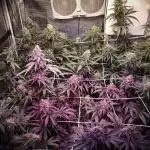
Week 22. Flowering4y ago


!["Jack Herer" C#4. Week 22 - Phi (/faɪ/;[1] uppercase Φ, lowercase φ or ϕ; Ancient Greek: ϕεῖ pheî [pʰéî̯]; M](https://bucket.growdiaries.com/static/post/photo/105536/6330531_grow-journal-by-exbeginner_m.jpg)


101.6 cm
Height
8 hrs
Light Schedule
19 °C
Day Air Temp
6.5
pH
Strong
Smell
1000 PPM
TDS
63 %
Air Humidity
19 °C
Solution Temp
19 °C
Substrate Temp
19 °C
Night Air Temp
26.5 l
Pot Size
45.72 cm
Lamp Distance
1250 PPM
CO₂ Level
Nutrients 1
Coconut Water
500 mll
Ultraviolet UV
3 likes
1 comment
Share
23
Week 23. Flowering4y ago


101.6 cm
Height
4 hrs
Light Schedule
16 °C
Day Air Temp
6.5
pH
Strong
Smell
1000 PPM
TDS
63 %
Air Humidity
16 °C
Solution Temp
16 °C
Substrate Temp
16 °C
Night Air Temp
100.31 l
Pot Size
45.72 cm
Lamp Distance
1250 PPM
CO₂ Level
Ultraviolet ggggg
4 likes
comments
Share
23
Week 23. Harvest4y ago


Spent 103 days
Ger Veg Flo Har
154 g
Bud dry weight per plant
Normal
Difficulty

Euphoric, Happy, Sleepy
Positive effects

Dry mouth
Negative effects

Depression, Insomnia, Stress
Medical effects

Earthy, Woody
Taste
Height
Day air temperature
Air humidity
PPM
PH
CO2
Light schedule
Solution temperature
Night air temperature
Substrate temperature
Pot size
Lamp distance


2 likes
2 comments
Share
Equipment Reviews


23
Week 23. Harvest4y ago
Happy Harvest Day!


8/10
Rated
Here we are once again, strong UV response can triple linalool content, this is the main terpene responsible for the amazing smell of GDP, and really makes for a uniquely strong-smelling perfection.
THC: 17%-23%
Δ9-THC: 13.98%
CBD:
Show more
Translate
Spent 103 days
Ger Veg Flo Har
83 g
Bud dry weight per plant
1
Plants
1.5 m²
Grow Room size
Easy
Difficulty

Relaxed, Sleepy, Uplifted
Positive effects
Dry mouth
Negative effects

Insomnia
Medical effects
Height
Day air temperature
Air humidity
PPM
PH
CO2
Light schedule
Solution temperature
Night air temperature
Substrate temperature
Pot size
Lamp distance


Ultraviolet tytyttyttytytytyt
2 likes
comments
Share
Equipment Reviews
23
Week 23. Harvest4y ago
Happy Harvest Day!

8/10
Rated
Jack Herer was a lifelong cannabis activist. He authored the non-fictional book The Emperor Wears No Clothes, which talks about the many beneficial uses of the plant for which he collected over years of compiling historical data.
The strain called Jack Herer is a tribute to this activist and was first cultivated under the care of Sensi Seeds. Its genetics stem from crossing Northern Lights #5, Shiva Skunk, and Haze making this hybrid ever popular for its appealing flavor and effects.
Once you open up a package of Jack Herer, you’ll be greeted with a light floral scent having undertones of fresh soil and lemon zest. The most notable characteristic about this strain is its vivid orange pistils scattered throughout light green flowers. A dusting of crystal trichomes gives it an even lighter appearance when it’s been cultivated properly.
Despite its odor being mild and its appearance seemingly like your average cannabis nug, reviewers say it’s ideal for cannabis consumers that are looking for a balance of effects. Jack Herer has been dubbed a “wake and bake” strain and is reputed to increase energy while relaxing the body just enough to want to keep moving. Users say this strain offers a mellow high accompanied with focus, so anyone pursuing creative tasks or conversations may benefit as cerebral activity enhances. Jack Herer has been acclaimed by users for improving visualization and brainstorming, while some have used it to reduce the symptoms of migraines, headaches, and stress. Be careful consuming it too late in the evening as it could keep you from falling asleep according to those who have tried this strain.
Overall, Jack Herer is a favorite for both newbies in the cannabis world as well as veteran consumers. Four phenotypes of this strain exist, making it one of the more diverse strains you’ll see on the market with two leaning towards indica traits and two leaning towards sativa traits.
Show more
Translate
Spent 103 days
Ger Veg Flo Har
180 g
Bud dry weight per plant
1
Plants
Height
Day air temperature
Air humidity
PPM
PH
CO2
Light schedule
Solution temperature
Night air temperature
Substrate temperature
Pot size
Lamp distance


Ultraviolet hhhhh
3 likes
comments
Share
Equipment Reviews
23
Week 23. Harvest4y ago
Happy Harvest Day!
8/10
Rated
Spent 103 days
Ger Veg Flo Har
350 g
Bud wet weight per plant
154 g
Bud dry weight per plant
1
Plants
Easy
Difficulty
Height
Day air temperature
Air humidity
PPM
PH
CO2
Light schedule
Solution temperature
Night air temperature
Substrate temperature
Pot size
Lamp distance


Ultraviolet gfgfggfgg
4 likes
comments
Share
Equipment Reviews
15 comments
Sort by
popularity
popularity
newest
oldest

homerjgangia commentedweek 04y ago
Good luck with your grow mate!💪💪
likes 6

THCanbisGrower commentedweek 04y ago
good luck with your grow man.
likes 5

Ultraviolet commented4y ago
@THCanbisGrower, Thank you brother!
likes 7

CannbellFarms commentedweek 224y ago
Best of luck Growing Brother! 🌱💪
likes 5

PuufPuuf commentedweek 114y ago
All the best to your new girls on thier journey to frostiness 🌱💚🍅
likes 5

StickyScissors commentedweek 164y ago
What are your thoughts on UVA. I have a 30W UVA but seldom turn it on. Kind of scares me 😞
likes 4
Ultraviolet commented4y ago
@StickyScissors, My thoughts are I wish id stop forgetting UV is on.........stoners beware!
likes 6
StickyScissors commented4y ago
@Ultraviolet, thank you! It’s scares me so I put it on a Wi-Fi enabled switch with a timer countdown. I like the colors they yield toward the end of flower.
likes 4

HAPPYWEEDS commentedweek 204y ago
You're doing good! Wish you all the best on your grow mate 😎
likes 4

MiyaguiOkPolilla commentedweek 194y ago
Vaya informacion tan valiosa amigo
likes 3
MiyaguiOkPolilla commented4y ago
@Ultraviolet, buenas respuestas crean sabiduria. Por que limitarnos? 🙏🍀👏
likes 2

BCbuds76 commentedweek 234y ago
Awesome diary!
likes 3
Ultraviolet commented4y ago
@BCbuds76, Thank you very much! I do try! I am forgetful in my old age it's good to keep Diaries!
likes 2

Natrona commentedweek 184y ago
What beauty!
likes 2
Natrona commentedweek 183y ago
👀 wowie,, I hope any of my ladies look as dazzling as this.
like 1

the end.
Enjoying this diary? Follow for more updates!

Prefer the old Diary view?
Go back to the old Diary view